Prostate Ultrasound and TRUS: What It Shows and Reading It
Reviewed by the LabReadAI medical team
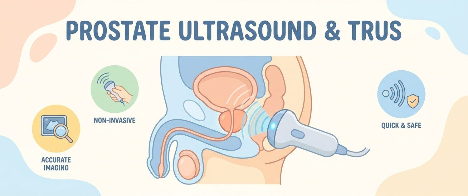
A prostate ultrasound and TRUS are often ordered for men with urinary complaints or with PSA deviations, and this raises questions: how these scans differ, what a prostate ultrasound shows, how to prepare for it and how to read the report. Let's go through it calmly and to the point, so you understand the meaning of each line in the protocol.
What a Prostate Ultrasound Shows
A prostate ultrasound assesses the gland with sound waves: its volume (size), shape, tissue structure, the presence of nodules and areas of altered density, and the state of the surrounding tissues. The bladder and the residual-urine volume are looked at separately. This scan does not make a diagnosis on its own, but it gives the doctor an important anatomical picture of the prostate.
Transabdominal and Transrectal Ultrasound
There are two main approaches. A transabdominal ultrasound is done with a probe over the front wall of the belly with a full bladder — it is less detailed but comfortable. A transrectal ultrasound (TRUS) is done with a thin probe through the rectum: it sits close to the prostate and gives a clearer image of the gland. Which option is needed is decided by the doctor for the task.
Why TRUS Is More Precise for the Prostate
TRUS is valued for detail: the probe's closeness to the gland lets it see the structure, small nodules and prostate zones better. That is why TRUS is used when precision matters — for example, with a raised PSA or suspicious changes. A transabdominal ultrasound more often serves for a general assessment of gland volume and residual urine.
Preparing for a Prostate Ultrasound
Preparation depends on the type of scan. For a transabdominal ultrasound you are usually asked to come with a full bladder (drink water and do not urinate before the procedure). TRUS may require emptying the bowel. Always check the exact requirements with the clinic in advance; the general principles are described in preparing for an ultrasound.
How to Read a Prostate Ultrasound Report
The protocol usually states the prostate volume (in cubic centimeters), the uniformity and echostructure of the tissue, the presence or absence of nodules, the state of the capsule and the residual-urine volume. An increased gland volume may accompany benign prostatic hyperplasia, and signs of inflammation — prostatitis. But an ultrasound report is a description, not a diagnosis: it is interpreted by a doctor together with the tests.
Prostate Ultrasound and PSA: How They Complement Each Other
Ultrasound and PSA solve different tasks and work as a pair. A PSA test is a lab marker of the gland's state, while ultrasound is its "picture": volume, structure, residual urine. Together they give the doctor a fuller picture than each alone, and help decide whether further tests are needed.
What to Do with the Report
Do not make a diagnosis from an ultrasound protocol yourself: the description of findings is assessed by a doctor in the context of complaints, PSA and examination. If the wording in the report is unclear, you can upload the scan for decoding — the service explains the terms in plain language and suggests questions to ask the urologist at the visit.
This article is for informational purposes only and does not replace a doctor's consultation. A prostate ultrasound and TRUS report is interpreted by a specialist in the context of the clinical picture.
For informational purposes only
This article is for informational purposes only and does not constitute medical advice, diagnosis, or treatment. Please consult a healthcare professional for medical guidance.