Uterine Fibroids: Types, Sizes, Symptoms and Pelvic Ultrasound
Reviewed by the LabReadAI medical team
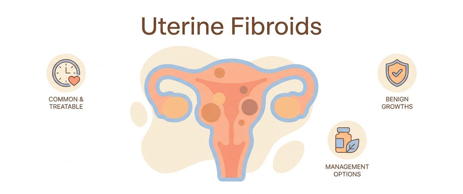
A diagnosis of "uterine fibroids" sounds frightening because of the word "tumor", but in fact it is one of the most common and almost always benign findings in women. Many live with fibroids for years without even knowing. Let's go through it calmly and by the facts: what uterine fibroids are, the types and sizes, which symptoms matter and what a pelvic ultrasound shows.
What Uterine Fibroids Are
Uterine fibroids are benign tumors made of the muscle tissue of the uterine wall (also called myomas or leiomyomas). They are not cancer and very rarely turn malignant. Fibroids are very common, especially at ages 30–50, and their growth is linked to female sex hormones: they often shrink after menopause. There may be one or several nodes of different sizes.
Types of Uterine Fibroids by Location
Fibroids are classified by where in the uterine wall they grow. A submucosal fibroid bulges into the uterine cavity and most often causes heavy bleeding. An intramural fibroid — the most common — sits within the wall. A subserosal fibroid grows outward, toward the abdominal cavity, and can press on neighboring organs. The type matters because symptoms and management depend on it.
Sizes of Uterine Fibroids
Node size is described in centimeters, and sometimes compared to weeks of pregnancy by the overall enlargement of the uterus. Small fibroids without symptoms are often simply watched. Large nodes or rapid growth need attention and a discussion of management with a doctor. The number of centimeters alone is not a verdict: what matters is the combination of size, location, symptoms and pregnancy plans.
Uterine Fibroid Symptoms
Fibroids are often symptomless and found by chance on ultrasound. When symptoms are present, the most common is heavy and prolonged periods, which can lead to anemia. There may also be a feeling of pressure and heaviness in the lower abdomen, frequent urination, discomfort during intimacy, less often pain. Cycle disturbances are covered in a separate article on cycle disruption and delay.
Pelvic Ultrasound for Fibroids
The main diagnostic method is a pelvic ultrasound. It shows the number of nodes, their sizes, location (submucosal, intramural, subserosal) and helps track fibroids over time. Ultrasound also tells fibroids from other lesions. If the report is unclear, you can upload the scan for decoding — the service explains the wording in plain language and suggests questions for your doctor.
Are Fibroids Dangerous and When to See a Doctor
Fibroids are benign, and most women do not need urgent intervention. See a gynecologist for heavy or prolonged periods, signs of anemia (weakness, pallor), pressure in the lower abdomen, rapid growth of a node or when planning a pregnancy. Similar symptoms are caused by other conditions — for example an ovarian cyst or endometriosis — so a doctor sorts it out.
Watch or Treat
Management depends not on the presence of fibroids but on symptoms, the size and location of nodes, age and pregnancy plans. Asymptomatic small fibroids are often simply watched on ultrasound, while active treatment is discussed for marked symptoms or large nodes. The decision on management is made by a doctor together with the patient, weighing benefits and risks.
This article is for informational purposes only and does not replace a doctor's consultation. The type and sizes of uterine fibroids and management are assessed by a specialist from the ultrasound and symptoms.
For informational purposes only
This article is for informational purposes only and does not constitute medical advice, diagnosis, or treatment. Please consult a healthcare professional for medical guidance.