Endometriosis: Symptoms, Diagnosis and Treatment Options
Reviewed by the LabReadAI medical team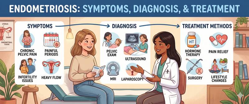
One in five women with endometriosis is told by a doctor that her pain is just "normal period pain" — and sent home with painkillers. On average, seven to ten years pass between the first symptoms and a confirmed diagnosis. During that time, endometriotic lesions grow, form adhesions and quietly erode fertility. Understanding what is happening in the body is the first step toward the right diagnosis.
What Endometriosis Is and How It Develops
Endometriosis is a chronic, hormone-dependent disease in which tissue resembling the endometrium — the inner lining of the uterus — is found and functions outside the uterine cavity. These lesions respond to cyclic oestrogen fluctuations: they swell in the luteal phase and "bleed" during menstruation — but the blood from ectopic deposits has nowhere to drain. It accumulates, triggers an inflammatory reaction, and leads to adhesions and "chocolate cysts" (endometriomas).
The most widely accepted theory of origin is retrograde menstruation: part of the menstrual blood carrying endometrial cells passes through the fallopian tubes into the peritoneal cavity. This occurs in most women, but a normally functioning immune system destroys these displaced cells. In endometriosis, something in this mechanism is defective: the cells implant, survive and proliferate. Genetic predisposition explains why first-degree female relatives of affected women face a seven- to tenfold higher risk.
The location of lesions determines the clinical picture. The most commonly affected sites are the ovaries (endometriomas), the rectouterine pouch, the uterosacral ligaments and the pelvic peritoneum. In deep infiltrating endometriosis (DIE), lesions penetrate the bladder, ureters, rectum and rectovaginal septum.
Staging by the rASRM classification (I–IV) reflects the extent and severity of adhesions but correlates poorly with pain. Clinically, it is more meaningful to distinguish between superficial peritoneal endometriosis, ovarian endometriomas and DIE — the last requiring a fundamentally different surgical approach.
Causes and Risk Factors
The precise cause of endometriosis is unknown. The contemporary model regards it as multifactorial — genetic susceptibility, immune dysregulation, hormonal imbalance and environmental factors all interact.
Genetics: a mother or sister with endometriosis increases risk seven- to tenfold. Several susceptibility loci have been identified but no single "endometriosis gene" exists.
Hormonal environment: endometriosis is oestrogen-dependent. Lesions contain excess aromatase enzyme, which synthesises oestradiol directly at the implantation site — local hyperoestrogenaemia sustains growth. Simultaneously, resistance to progesterone is impaired.
Immune dysfunction: peritoneal NK cells and macrophages are less effective at eliminating ectopic endometrial cells. Pro-inflammatory cytokines in the peritoneal fluid create conditions favourable for implantation and growth.
Factors that increase risk: early menarche (before 12), short cycles (< 27 days), heavy periods, nulliparity, low BMI.
Factors that reduce risk: pregnancies, lactation, long-term oral contraceptive use, regular physical exercise.
An important distinction: polycystic ovary syndrome and endometriosis are not the same condition, though both impair reproductive function. PCOS is characterised by androgen excess and anovulation; endometriosis by hyperoestrogenaemia and pain. In some women both coexist, substantially complicating diagnosis.
Symptoms: Pain and Infertility
The two hallmarks of endometriosis are chronic pelvic pain and reduced fertility. Their severity does not correlate with disease stage.
Pain:
- Dysmenorrhoea — painful periods not controlled by standard NSAIDs. Periods that are disabling for one to two days a month require investigation, not simply painkillers
- Chronic pelvic pain — dull or cramping pain in the lower abdomen or spine persisting outside menstruation for more than 6 months
- Dyspareunia — painful intercourse with deep penetration. Classically associated with endometriosis of the posterior Douglas pouch
- Dyschezia — painful defecation worsening during menstruation. Characteristic of rectovaginal endometriosis
- Dysuria — painful urination intensifying before menstruation
Menstrual disturbances: heavy periods (menorrhagia), intermenstrual spotting, premenstrual brown discharge.
Infertility: endometriosis is found in 25–50% of women presenting with infertility. Adhesions distort the fallopian tubes; endometriomas reduce ovarian reserve; the peritoneal inflammatory milieu is toxic to sperm and embryos; immune abnormalities interfere with implantation.
Diagnosis: Laparoscopy, Ultrasound and Blood Tests
Laparoscopy with biopsy and histological confirmation remains the gold standard. However, current guidelines support a clinical diagnosis when history and ultrasound findings are characteristic — treatment may be started without surgery.
Transvaginal ultrasound — the first-line investigation. Sensitivity for ovarian endometriomas is 90–95%: the characteristic appearance is a unilocular cyst with homogeneous ground-glass echogenicity. MRI of the pelvis is best for mapping DIE involving the bowel and urinary tract.
Laparoscopy with biopsy — mandatory for atypical presentations, suspected DIE, or failure of hormonal therapy.
Laboratory tests:
CA-125 — elevated in 75–80% of stage III–IV patients but only 20–25% at stage I–II. The main value is monitoring: a rising level after surgery or during treatment signals progression. CA-125 above 200 U/mL in an endometrioma requires extended oncological evaluation with HE4 and the ROMA index.
Sex hormone panel — FSH, LH, oestradiol, AMH. AMH is critical: endometriomas reduce ovarian reserve, and AMH below 1.0 ng/mL before surgery signals risk of further depletion. This determines the approach for women planning pregnancy.
Complete blood count — heavy periods frequently cause iron deficiency anaemia: recurrent large blood loss depletes iron stores progressively.
Endometriosis and the Risk of Ovarian Cancer
The link between endometriosis and ovarian cancer is real and clinically significant: women with endometriosis have approximately three times the population risk of ovarian cancer. But the absolute risk remains low — the vast majority of women with endometriosis never develop cancer.
The histological subtypes most closely associated with endometriosis are clear cell carcinoma and endometrioid carcinoma. The mechanism is malignant transformation of endometrioma cells driven by chronic oxidative stress and inflammation.
Features associated with elevated transformation risk: disease duration above 10 years, large endometriomas (> 9 cm), growth of the cyst or new solid component on follow-up imaging, age over 45, postmenopausal status. When these features are present, ultrasound intervals are shortened to 3 months and HE4 with ROMA index calculation is added.
Treatment: Hormones, Surgery and Pain Management
Treating endometriosis is a long-term strategy. Three factors determine the approach: severity of pain, desire to preserve fertility, and stage and location of lesions.
Hormonal therapy:
Combined oral contraceptives (COCs) — first-line for dysmenorrhoea and chronic pelvic pain. A continuous regimen (no placebo interval) is more effective than cyclic dosing.
Progestins (dienogest, medroxyprogesterone) — as effective as COCs for pain; dienogest is superior for deep endometriosis and reduces endometrioma size with prolonged use.
GnRH agonists (leuprorelin, buserelin) — induce medical menopause. Effective for severe pain and IVF preparation. Mandatory "add-back" low-dose oestrogen-progestin therapy protects bone density; without it, treatment is limited to 6 months.
Surgical treatment:
Laparoscopic lesion excision is preferred over coagulation: it provides histological confirmation and lower recurrence risk. For endometriomas: cystectomy reduces recurrence but irreversibly reduces ovarian reserve. AMH must be measured before ovarian surgery in women planning pregnancy.
DIE involving the bowel or urinary tract requires surgery in a specialist endometriosis centre with a gynaecologist, colorectal surgeon and urologist.
Fertility treatment: stage I–II with patent tubes — ovulation induction with or without IUI. Stage III–IV or previous treatment failure — IVF.
When to See a Doctor
Painful periods that interfere with daily life are not a normal variant and require investigation, not simply analgesics.
See a gynaecologist when one or more of the following apply:
- dysmenorrhoea severe enough to require prescription pain relief or to miss work or studies;
- chronic pelvic pain outside menstruation lasting more than 3 months;
- deep dyspareunia;
- difficulty conceiving after 6–12 months of regular unprotected intercourse;
- an ovarian cyst with endometrioma characteristics found incidentally;
- a mother or sister has been diagnosed with endometriosis.
Endometriosis is a chronic condition without a definitive cure, but effective symptom control and fertility preservation are achievable. Early diagnosis and a long-term treatment strategy agreed with a specialist are the keys to a better prognosis.
This content is for informational purposes only and does not replace professional medical advice.
For informational purposes only
This article is for informational purposes only and does not constitute medical advice, diagnosis, or treatment. Please consult a healthcare professional for medical guidance.