ACTH Test: Normal Range, High and Low Levels, Interpretation
Reviewed by the LabReadAI medical team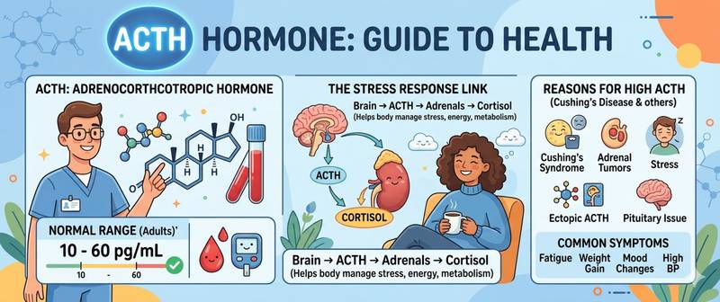
ACTH is a hormone that does not work alone. It is meaningless without cortisol and uninterpretable without it. ACTH is almost never ordered in isolation: its diagnostic value emerges only in combination with cortisol — and it is their ratio that distinguishes a pituitary tumor from an adrenal one, primary adrenal insufficiency from secondary, and functional stress from true pathology. Let's break down how this hormone works, how to test it correctly, and how to read the result.
What ACTH Is and How the HPA Axis Works
ACTH (adrenocorticotropic hormone, corticotropin) is a 39-amino-acid peptide hormone synthesized by the anterior pituitary gland. Its job is to stimulate the adrenal cortex to produce cortisol, and to a lesser extent androgens and mineralocorticoids.
ACTH is the central element of the hypothalamic-pituitary-adrenal (HPA) axis:
- The hypothalamus releases corticotropin-releasing hormone (CRH)
- The pituitary secretes ACTH into the bloodstream in response to CRH
- The adrenal cortex synthesizes cortisol under ACTH stimulation
- Cortisol suppresses CRH and ACTH through negative feedback
This closed loop maintains cortisol within the physiological range. Failure at any link — hypothalamus, pituitary, or adrenal — disrupts the entire system, and the nature of the failure is determined precisely by the ACTH-to-cortisol ratio.
ACTH secretion has a pronounced circadian rhythm: peak in the morning (4–8 AM), nadir late at night (10 PM–midnight). The morning ACTH surge triggers the morning cortisol peak — this is the biological basis of waking. Acute stress (physical, emotional, hypoglycemia, fever, surgery) can elevate ACTH 5–10-fold within minutes — an evolutionary mobilization mechanism.
Beyond cortisol stimulation, ACTH has a mild effect on DHEA-S synthesis — the principal adrenal androgen. This is why ACTH-deficient states gradually deplete both cortisol and DHEA-S.
Normal ACTH Range
ACTH is unstable in plasma — it degrades rapidly outside the body. This makes the assay highly demanding pre-analytically. Reference ranges depend on collection time and vary slightly between laboratories.
| Collection time | ACTH normal range (pg/mL) |
|---|---|
| Morning (7:00–10:00 AM) | 7.2–63 |
| Evening (6:00–10:00 PM) | < 30 |
| Late night (11 PM–1 AM) | < 10 |
Children have higher normal ranges; in older adults the lower bound may shift to 5 pg/mL. Pregnancy physiologically raises ACTH 1.5–2-fold in the second and third trimesters.
Unit conversion: pg/mL × 0.22 = pmol/L.
Critically: ACTH is interpreted not as a standalone number but in the context of simultaneously measured cortisol — see the section below.
How to Prepare for an ACTH Test
ACTH testing is among the most pre-analytically challenging assays. Failure to follow conditions can fully invalidate the result.
Preparation and collection:
- Strictly fasting, drawn at 8:00–10:00 AM — at the peak of the circadian rhythm
- Avoid intense exercise and emotional stress for 24 hours beforehand
- No smoking for at least 3 hours before the draw
- Blood is collected into a chilled EDTA tube, immediately centrifuged, and frozen — otherwise ACTH degrades within 10–15 minutes at room temperature
- Disclose to the lab any glucocorticoids (prednisolone, dexamethasone, hydrocortisone), estrogens, or amphetamines — all alter ACTH
- When Addison's disease is suspected — a repeat draw at 4:00–6:00 PM; in this case, high ACTH with low cortisol confirms the diagnosis
Indications:
- Suspected Cushing's syndrome (paired with cortisol and DHEA-S)
- Suspected Addison's disease or secondary adrenal insufficiency
- Differential diagnosis of hypercortisolism causes
- Monitoring of pituitary or adrenal tumor therapy
- Stimulation tests (with CRH) and suppression tests (dexamethasone)
For comprehensive HPA-axis assessment, ACTH is frequently included in a hormone panel alongside cortisol, TSH, and prolactin.
Causes of High ACTH
Elevated ACTH signals that the pituitary is straining to "reach" the adrenal glands. This occurs either with autonomous pituitary pathology or with adrenal insufficiency.
| Cause | Cortisol | ACTH | Mechanism |
|---|---|---|---|
| Cushing's disease (ACTH-secreting pituitary adenoma) | High | High or upper-normal | Autonomous ACTH secretion by a pituitary tumor |
| Ectopic ACTH syndrome (small-cell lung cancer, carcinoid) | Very high | Very high (> 200 pg/mL) | Non-pituitary tumor produces ACTH |
| Addison's disease (primary AI) | Low | Very high | Pituitary attempting to stimulate destroyed adrenals |
| Congenital adrenal hyperplasia | Normal or low | High | Steroidogenic enzyme deficiency |
| Chronic stress, depression | Normal or modestly elevated | Modestly elevated | Functional HPA hyperactivation |
| Hypothalamic CRH-secreting tumor | High | High | Tertiary hypercortisolism |
| Abrupt glucocorticoid withdrawal after long-term use | Low (atrophy) | Gradually rising | Pituitary disinhibition with persistent adrenal atrophy |
Causes of Low ACTH
Low ACTH means the pituitary is unable or not required to stimulate the adrenals. This occurs either with autonomous cortisol production outside pituitary control or with pituitary damage itself.
| Cause | Cortisol | ACTH | Mechanism |
|---|---|---|---|
| Adrenal cortical adenoma (corticosteroma) | High | Suppressed (< 5 pg/mL) | Autonomous cortisol suppresses pituitary |
| Long-term glucocorticoid use | Low endogenous | Very low | Exogenous cortisol suppresses HPA axis |
| Secondary adrenal insufficiency | Low | Low or normal (inappropriately low) | Pituitary lesion — no adrenal stimulation |
| Sheehan's syndrome (pituitary necrosis) | Low | Low | Postpartum pituitary failure |
| Craniopharyngioma, mass-effect pituitary adenoma | Low | Low | Mechanical destruction of ACTH-secreting cells |
| Head trauma, pituitary radiotherapy | Low | Low | Structural pituitary damage |
ACTH and Cortisol: Four Interpretation Scenarios
Without simultaneous cortisol assessment, an ACTH result is half a diagnosis. All clinically relevant scenarios reduce to four combinations:
High cortisol + high ACTH → ACTH-dependent hypercortisolism. The source is the pituitary (Cushing's disease, ~80% of cases) or an ectopic tumor (~20%). Differentiated by the high-dose dexamethasone suppression test and pituitary MRI.
High cortisol + low ACTH (< 5 pg/mL) → ACTH-independent hypercortisolism. The source is in the adrenal: adenoma, carcinoma, less commonly bilateral hyperplasia. Adrenal CT or MRI is mandatory.
Low cortisol + high ACTH → primary adrenal insufficiency (Addison's disease). The pituitary is intact; the adrenals themselves are destroyed — autoimmune, infectious, or metastatic. Confirmed by the tetracosactide (Synacthen) stimulation test.
Low cortisol + low ACTH → secondary or tertiary adrenal insufficiency. The pituitary or hypothalamus is damaged. Most often a consequence of long-term glucocorticoid therapy; less commonly pituitary adenoma, Sheehan's syndrome, or head trauma.
This four-quadrant algorithm is the foundation of all adrenal diagnostics and should be familiar to anyone holding their ACTH result.
Dexamethasone Suppression and Stimulation Tests
Standalone ACTH measurement is often insufficient — particularly at borderline values. Functional tests fill this gap.
Low-dose dexamethasone suppression test (overnight): the patient takes 1 mg dexamethasone at 11 PM; cortisol and ACTH are drawn at 8 AM. In a healthy person, dexamethasone suppresses ACTH via negative feedback, and morning cortisol falls below 50 nmol/L. Failure to suppress is a screening signal for Cushing's syndrome.
High-dose dexamethasone suppression test (8 mg over 2 days): differentiates Cushing's disease from ectopic ACTH secretion. A pituitary adenoma is partially suppressed at high doses; an ectopic tumor is not.
Tetracosactide (Synacthen) stimulation test — for suspected adrenal insufficiency: synthetic ACTH is administered, and cortisol is measured 30–60 minutes later. An adequate rise (> 500 nmol/L) excludes primary AI.
These tests are interpreted by an endocrinologist — self-interpretation is unproductive.
When to See an Endocrinologist
Any non-physiological ACTH result is grounds for an endocrinology referral. Practical thresholds:
- ACTH < 5 pg/mL with elevated cortisol — mandatory exclusion of an adrenal tumor (CT/MRI)
- ACTH > 100 pg/mL — mandatory exclusion of an ACTH-secreting tumor (contrast pituitary MRI; chest and abdominal CT if ectopic secretion is suspected)
- High ACTH with low cortisol — suspected Addison's disease; urgent: 21-hydroxylase antibodies, electrolytes, glucose
- Any patient on a long glucocorticoid course with fatigue, hypotension, or nausea — assess ACTH and cortisol before discontinuation
Emergency care is required for the combination of hypotension, nausea, vomiting, hypoglycemia, and altered consciousness: an Addisonian crisis is possible, and intravenous hydrocortisone is needed without delay.
For comprehensive adrenal function assessment, the optimal starting point is the adrenal stress panel, which includes ACTH, morning and evening cortisol, DHEA-S, and prolactin. For DHEA-S as a parallel marker of adrenal function and its role in longevity, see the dedicated article on DHEA-S and aging.
This article is for informational purposes only and does not replace professional medical advice. ACTH interpretation requires simultaneous cortisol assessment and functional testing under endocrinologist supervision.
For informational purposes only
This article is for informational purposes only and does not constitute medical advice, diagnosis, or treatment. Please consult a healthcare professional for medical guidance.