Cortisol Blood Test: Normal Levels, Causes and Interpretation
Reviewed by the LabReadAI medical team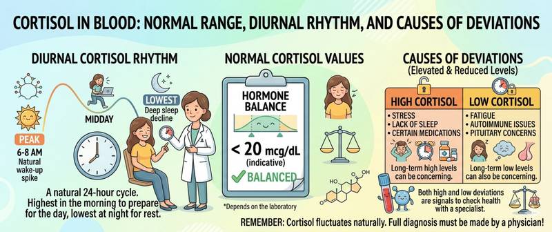
Cortisol is a hormone that simultaneously saves lives and undermines health. In an acute situation it is indispensable: it mobilizes energy, suppresses inflammation, and maintains blood pressure. But in chronic excess it begins doing the very things it is meant to prevent — breaking down muscle, raising blood pressure, impairing immunity, and depositing fat on the abdomen. Let's break down its biochemistry, normal ranges, and why the time of testing is fundamentally important.
What Cortisol Is and How the HPA Axis Works
Cortisol is a steroid hormone of the glucocorticoid class, synthesized in the adrenal cortex (zona fasciculata). It is regulated through a three-tier hierarchy — the hypothalamic-pituitary-adrenal (HPA) axis:
- The hypothalamus releases corticotropin-releasing hormone (CRH) → stimulates the pituitary
- The pituitary releases ACTH (adrenocorticotropic hormone) → stimulates the adrenal cortex
- The adrenal glands synthesize cortisol → through negative feedback, suppress CRH and ACTH
The defining characteristic of cortisol is its pronounced circadian rhythm: concentrations are approximately 8-fold higher in the morning (peak at 6–8 AM) than late at night. This rhythm governs not only biological processes but the entire logic of diagnostics: the same value of 400 nmol/L means "normal" in the morning and "significantly elevated" late at night.
Physiological functions of cortisol:
- Metabolic regulation — raises blood glucose through gluconeogenesis; stimulates protein and fat breakdown as energy substrates
- Anti-inflammatory action — suppresses pro-inflammatory cytokine synthesis and stabilizes immune cell membranes; this is the basis for synthetic glucocorticoid use in medicine
- Vascular tone — maintains vascular sensitivity to catecholamines; in adrenal insufficiency, hypotension becomes resistant to vasopressors
- Sodium retention — a mild mineralocorticoid effect; in excess (Cushing's syndrome) — hypernatremia and hypokalemia
- Immunomodulation — suppresses cellular immunity; chronically high cortisol reduces NK-cell and T-lymphocyte activity
A practical guide to reducing chronically elevated cortisol in women is in the article how to lower cortisol in women.
Normal Cortisol Levels: Blood, Saliva and Urine
Cortisol reference values critically depend on the time of day and the biological specimen. Without documentation of collection time, any cortisol value is uninterpretable.
Blood cortisol (serum/plasma):
| Collection time | Normal (nmol/L) | Normal (µg/dL) |
|---|---|---|
| Morning 8:00–10:00 AM (peak) | 138–690 | 5–25 |
| Midday 12:00–4:00 PM | 83–413 | 3–15 |
| Evening 4:00–8:00 PM | 69–345 | 2.5–12.5 |
| Late night 11:00 PM–midnight | < 50 | < 1.8 |
Unit conversion: nmol/L ÷ 27.59 = µg/dL.
Salivary cortisol (preferred for diurnal profiling and Cushing's syndrome screening):
| Time | Normal (nmol/L) |
|---|---|
| Morning (30 min after waking) | 10–45 |
| Midday | 2–11 |
| Late evening (10:00–11:00 PM) | < 4.3 nmol/L |
Late-night salivary cortisol < 4.3 nmol/L is the most sensitive single screening test for Cushing's syndrome.
Free cortisol in 24-hour urine (reflects total daily cortisol output):
| Group | Normal (nmol/24h) | Normal (µg/24h) |
|---|---|---|
| Adults | 55–330 | 20–120 |
During pregnancy, urinary free cortisol physiologically rises 2–3-fold — interpret with caution.
How to Prepare for a Cortisol Blood Test
Cortisol testing is one of the most pre-analytically demanding hormonal assays. Failure to follow conditions can completely invalidate the result.
Blood cortisol:
- Strictly fasting, drawn at 8:00–10:00 AM — this is when cortisol is at its physiological peak; an afternoon draw produces a physiologically low result easily mistaken for pathology
- Avoid intense physical exercise and emotional stress for 24 hours beforehand — both powerfully activate the HPA axis
- Blood is drawn after the patient has rested quietly for 30 minutes — venipuncture itself raises cortisol
- When Cushing's syndrome is suspected — a late-night cortisol (11:00 PM) is also collected: elevation above 207 nmol/L is a diagnostic criterion for autonomous cortisol excess
- Disclose all glucocorticoid use (prednisolone, dexamethasone, budesonide, even topical steroids with systemic absorption) — they suppress cortisol through HPA axis suppression
Low-dose dexamethasone suppression test: The patient takes 1 mg dexamethasone at 11:00 PM; cortisol is measured at 8:00 AM. With normal HPA axis function: suppression to < 50 nmol/L. Failure to suppress is a Cushing's syndrome screening signal.
The relationship with other hormones: TSH and cortisol are often measured together when patients complain of fatigue and weight changes — they mutually influence thyroid and adrenal function.
Causes of High Cortisol (Hypercortisolism)
Hypercortisolism is divided into endogenous (excess production within the body) and exogenous (glucocorticoid medication use).
| Cause | Mechanism | ACTH level | Characteristic features |
|---|---|---|---|
| Cushing's disease (pituitary ACTH adenoma) | Autonomous ACTH → adrenal stimulation | High | Central obesity; moon face; striae |
| Cushing's syndrome (adrenal adenoma) | Autonomous adrenal cortisol | Low (suppressed) | Same features; unilateral adrenal mass |
| Ectopic ACTH syndrome (lung, pancreatic tumors) | Tumor produces ACTH outside the pituitary | Very high | Rapid onset; hypokalemia; hyperpigmentation |
| Exogenous glucocorticoids | External GC administration | Very low (suppressed) | Clear link to medication |
| Chronic psychoemotional stress | HPA axis activation | Modestly elevated | Unstable moderate elevation |
| Obesity | Impaired HPA feedback | Normal | Moderate elevation; no pathological Cushing's stigmata |
| Depression and anxiety disorders | Hypothalamic hyperactivation | Modestly elevated | Psychiatric symptoms |
| Alcohol use disorder (pseudo-Cushing's) | Alcohol-driven HPA stimulation | Variable | Cushing's-like features; normalizes with abstinence |
Clinical signs of chronic hypercortisolism:
- Central obesity (moon face, dorsocervical fat pad, thin limbs)
- Wide (> 1 cm) purple-red striae on abdomen, thighs, and shoulders
- Proximal muscle weakness (difficulty rising from a chair without arm support)
- Hypertension resistant to treatment
- Diabetes or impaired glucose tolerance
- Osteoporosis
- Depression and cognitive impairment
Even moderate sustained hypercortisolism suppresses prolactin and sex hormones through inhibitory effects on GnRH — explaining menstrual irregularities and reduced libido during chronic stress.
Causes of Low Cortisol (Hypocortisolism)
Reduced cortisol — adrenal insufficiency — is less common but potentially life-threatening.
| Cause | Mechanism | Characteristic features |
|---|---|---|
| Addison's disease (primary AI) | Autoimmune or infectious destruction of adrenal cortex | Hyperpigmentation; hypotension; hyponatremia + hyperkalemia |
| Secondary AI (pituitary pathology) | ACTH deficiency → adrenal atrophy | Pallor (no ACTH → no skin pigmentation) |
| Tertiary AI (hypothalamic pathology) | CRH deficiency → ACTH deficiency | Trauma, hypothalamic tumors |
| Glucocorticoid withdrawal | Long-term GCs suppressed HPA axis → adrenal atrophy | Abrupt cessation after prolonged GC course |
| Sheehan's syndrome | Pituitary necrosis from postpartum hemorrhage | Agalactia; amenorrhea |
| Tuberculous adrenalitis | Adrenal cortex destruction | Adrenal calcifications on CT |
Addisonian crisis — acute adrenal insufficiency — is a rare but real medical emergency: sudden severe hypotension, circulatory collapse, hypoglycemia, hyperkalemia, vomiting, and loss of consciousness. Precipitated by stress, infection, or abrupt glucocorticoid withdrawal in a patient with chronic AI.
In hypocortisolism, calcitriol synthesis is impaired and calcium absorption falls — linking cortisol deficiency to vitamin D and calcium deficiency in patients with chronic adrenal insufficiency.
Cortisol and Other Hormones: Systemic Connections
Cortisol is the "conductor" of the stress response — its deviations immediately ripple through the entire endocrine system.
Cortisol and insulin. Cortisol is a physiological insulin antagonist: it raises blood glucose and reduces tissue insulin sensitivity. Chronic hypercortisolism is a recognized cause of secondary diabetes. Insulin resistance in metabolic syndrome is partly driven by heightened HPA axis activity.
Cortisol and TSH. Cortisol impairs T4-to-T3 conversion and reduces pituitary sensitivity to TRH. This explains why patients with chronic stress frequently have hypothyroid-like symptoms with a technically normal TSH — the so-called "normal TSH with peripheral thyroid dysfunction" pattern.
Cortisol and sodium. Cortisol exerts a mild mineralocorticoid effect, retaining sodium and water. In Addison's disease the opposite occurs: hyponatremia and hyperkalemia, from absence of both cortisol and aldosterone.
Cortisol and prolactin. Acute stress simultaneously raises both hormones — through shared neuroendocrine pathways. Chronically elevated cortisol, however, suppresses prolactin via dopaminergic mechanisms. Distinguishing these patterns is important when evaluating hyperprolactinemia in chronically stressed patients.
Cortisol and sex hormones. "Progesterone steal": under chronic stress, pregnenolone — the shared precursor of all steroid hormones — is preferentially channeled toward cortisol synthesis at the expense of sex hormones. This is one mechanism underlying cycle disruption, infertility, and reduced libido under chronic stress.
When Cortisol Abnormalities Require Medical Attention
Scheduled visit to an endocrinologist when:
- Morning cortisol > 690 nmol/L on two independent measurements with proper collection conditions — Cushing's syndrome screening
- Positive dexamethasone suppression test (failure to suppress below 50 nmol/L) — pituitary MRI and/or adrenal CT is mandatory
- Morning cortisol < 138 nmol/L on two measurements — exclude adrenal insufficiency; Synacthen stimulation test
- Clinical signs of hyper- or hypocortisolism at any cortisol level — the clinical picture matters more than the number
Call emergency services immediately when:
- Sudden severe hypotension (BP < 90/60 mmHg) combined with nausea, vomiting, and abdominal pain in a patient with known AI or who has taken glucocorticoids — possible Addisonian crisis
- Impaired consciousness, seizures, and hypoglycemia in a patient with AI — intravenous hydrocortisone must be given immediately
Chronically elevated cortisol is a key driver of accelerated biological ageing — triggering insulin resistance, inflammation and muscle loss. The cellular mechanisms behind this are covered in the article ageing of the body: causes and mechanisms, and a practical longevity programme is in the article how to live long and healthy. For the cortisol–DHEA-S axis and adrenal hormone balance in the longevity context, see the article DHEA-S and aging.
This article is for informational purposes only and does not replace professional medical advice. Consult an endocrinologist if your cortisol level is outside the normal range.
For informational purposes only
This article is for informational purposes only and does not constitute medical advice, diagnosis, or treatment. Please consult a healthcare professional for medical guidance.