Rheumatoid Factor (RF): Normal Range, Results and Causes
Reviewed by the LabReadAI medical team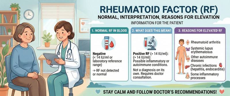
You get your blood test results back and spot "rheumatoid factor" flagged as high. The word "autoimmune" is already in your search bar — and the worry sets in quickly. Don't jump to conclusions: an elevated RF does not always mean disease, and in many cases this value rises even in completely healthy people. This article covers what rheumatoid factor is, the reference ranges, what a high result actually indicates, and when you need to see a rheumatologist urgently.
What Is Rheumatoid Factor and Why Is It Tested
Think of the immune system as a precisely calibrated radar: in health it targets foreign threats while leaving the body's own tissue untouched. In some conditions this calibration fails — and the radar starts firing at friendly targets. Rheumatoid factor (RF) consists of IgM antibodies that the immune system mistakenly produces against its own IgG immunoglobulins. In effect, one class of protective proteins attacks another.
RF was first described in 1940 by Norwegian researcher Erik Waaler and has since become one of the core assays in rheumatology and immunology.
The test is ordered when a patient has: joint pain, swelling, or stiffness — particularly on waking; symmetrical involvement of small hand and foot joints; signs of systemic inflammation (fever, fatigue, weight loss); or suspected connective tissue autoimmune disease. RF is often ordered alongside a complete blood count to assess leukocyte and platelet levels and check for anaemia, which frequently accompanies inflammatory conditions.
A positive RF most commonly points to rheumatoid arthritis — the most prevalent chronic inflammatory joint disease. However, it is not a definitive diagnostic test: the result must always be interpreted in the context of clinical findings and other investigations.
RF is not a population screening test. Ordering it in people without joint symptoms leads to a high rate of false-positive results and unnecessary anxiety.
How to Prepare for a Rheumatoid Factor Test
Blood is drawn from a vein, typically the antecubital fossa. Preparation is straightforward, but a few steps matter for an accurate result.
When to go: ideally in the morning, between 7 and 11 am. Fast for at least 8–12 hours beforehand — a full meal can affect the rheological properties of serum and reduce the accuracy of immunological tests. Drinking water is fine.
24 hours before the test: avoid strenuous exercise and alcohol. If you are acutely unwell or have been vaccinated within the past 2–3 weeks, let your doctor know — this can temporarily elevate the result.
Medications: certain immunosuppressants (methotrexate, prednisolone, hydroxychloroquine) and NSAIDs can lower RF levels. Do not stop any medication without medical advice — ask your doctor whether a temporary pause is needed before testing.
Testing methods: modern laboratories use latex agglutination (semi-quantitative), nephelometry, immunoturbidimetry (both quantitative), and ELISA. Different methods use different units and cutoff values, which is why results from different laboratories should not be directly compared without checking the method used. Results are typically available within 1–2 business days.
Rheumatoid Factor Normal Range: Reference Values
Most laboratories apply a single reference range for all adults regardless of sex. Minor differences between labs reflect different methods and reagents — always use the range printed on your own report.
| Group | RF Normal Range | Notes |
|---|---|---|
| Adults 18–65 | < 14 IU/mL | Sex-independent |
| Children under 12 | < 12.5 IU/mL | Rarely tested in paediatrics |
| Adults over 65 | Up to 20 IU/mL | Mild elevation may be physiologically normal |
The clinical significance of an elevated RF is graded by level:
- 14–50 IU/mL — weakly positive. May be physiological or infection-related; needs clarification.
- 50–100 IU/mL — moderately positive. Raises suspicion of autoimmune inflammation; comprehensive workup indicated.
- > 100 IU/mL — strongly positive. Significant autoimmune involvement, most often rheumatoid arthritis or another systemic disease.
The reference range is a probability boundary, not an absolute threshold. A value of 15 IU/mL with no symptoms requires no treatment; 12 IU/mL in the setting of active synovitis absolutely does.
Causes of Elevated Rheumatoid Factor
An elevated RF is a signal for investigation, not a diagnosis. Causes fall into two distinct categories.
Autoimmune and Inflammatory Conditions
The most common and clinically significant cause is rheumatoid arthritis: 70–80% of patients test positive, even in early disease. High RF in RA correlates with a more aggressive disease course and a greater risk of joint destruction.
Other conditions associated with elevated RF include Sjögren's syndrome (75–95% positivity — among the highest rates of any condition), systemic lupus erythematosus, mixed connective tissue disease, systemic sclerosis, dermatomyositis, and antiphospholipid syndrome.
Autoimmune activity is consistently confirmed by a parallel rise in C-reactive protein — an acute-phase marker that reflects the intensity of the current inflammatory process.
Non-Autoimmune Causes
Chronic infections can produce moderate or even significant RF elevation. This is not an immune error but a normal response to prolonged antigenic stimulation. The most frequent infectious causes are viral hepatitis B and C, tuberculosis, infective endocarditis, and chronic Lyme disease.
Non-infectious, non-autoimmune causes include chronic lung disease (chronic bronchitis, sarcoidosis), liver cirrhosis, and haematological malignancies (lymphomas, chronic lymphocytic leukaemia).
ESR is also elevated in these settings, and comparing it with RF and C-reactive protein helps clinicians decide which diagnostic direction to pursue.
Negative Rheumatoid Factor: Seronegative Arthritis
In 20–30% of patients with confirmed rheumatoid arthritis, RF remains within the normal range. This is called seronegative disease — it follows the same clinical course as classic RA but is invisible to the RF test.
The underlying reason is often genetic: certain HLA-DRB1 gene variants are associated with a tendency toward seronegative disease. In these patients joint inflammation can be just as active as in seropositive RA, and in some cases even more destructive.
This is where anti-cyclic citrullinated peptide antibodies (anti-CCP) become critical. The test is considerably more specific for rheumatoid arthritis and can detect the disease in seronegative cases. In some patients, anti-CCP turns positive years before clinical symptoms appear — allowing early intervention.
A negative RF with no symptoms is simply normal. If joint symptoms are present, a normal result does not exclude disease, and further diagnostic workup should continue.
False-Positive RF: When the Result Does Not Mean Arthritis
False-positive results are one of the main sources of anxiety for patients who discover elevated RF during a routine health screen.
Age. In people over 65, a mild elevation (up to 15–25 IU/mL) occurs in 10–25% of individuals without any underlying disease. This reflects normal age-related changes in immune regulation.
Recent infections and vaccination. An acute viral illness or vaccination within 2–3 weeks before testing temporarily activates the immune system and can produce a mild RF elevation, which typically resolves after recovery.
Pregnancy. Normal immune changes during pregnancy are sometimes reflected in false-positive immunological tests, including RF.
Other chronic conditions. Haematological malignancies, primary biliary cirrhosis, and sarcoidosis can all raise RF without any joint involvement.
A single elevated RF cannot support a diagnosis. The clinician will always correlate the result with symptoms, physical examination findings, additional blood tests, and imaging.
When to See a Doctor
Prompt evaluation by a GP or rheumatologist is warranted when:
- RF exceeds the upper limit of normal by a factor of 3 or more (> 42 IU/mL when the cutoff is 14 IU/mL).
- Elevated RF is accompanied by joint swelling, pain, and morning stiffness lasting more than 30–60 minutes.
- C-reactive protein and ESR are simultaneously elevated — a pattern of active systemic inflammation requiring prompt diagnosis.
- Symptoms extend beyond joints: persistent dry eyes and mouth, a butterfly-shaped facial rash, hair loss, or unrelenting fatigue and weight loss.
- Several small joints of the hands or feet are symmetrically affected — particularly in women aged 30–50.
Early treatment is critical: for rheumatoid arthritis, therapy initiated within the first 3–6 months of symptom onset can prevent irreversible cartilage and bone destruction. Delaying by 6–12 months significantly worsens long-term prognosis.
This article is for informational purposes only and does not replace medical consultation. Result interpretation and diagnosis are made by a qualified physician.
For informational purposes only
This article is for informational purposes only and does not constitute medical advice, diagnosis, or treatment. Please consult a healthcare professional for medical guidance.