How to Open a DICOM (.dcm) File: Free Viewers and Online Options
Reviewed by the LabReadAI medical team
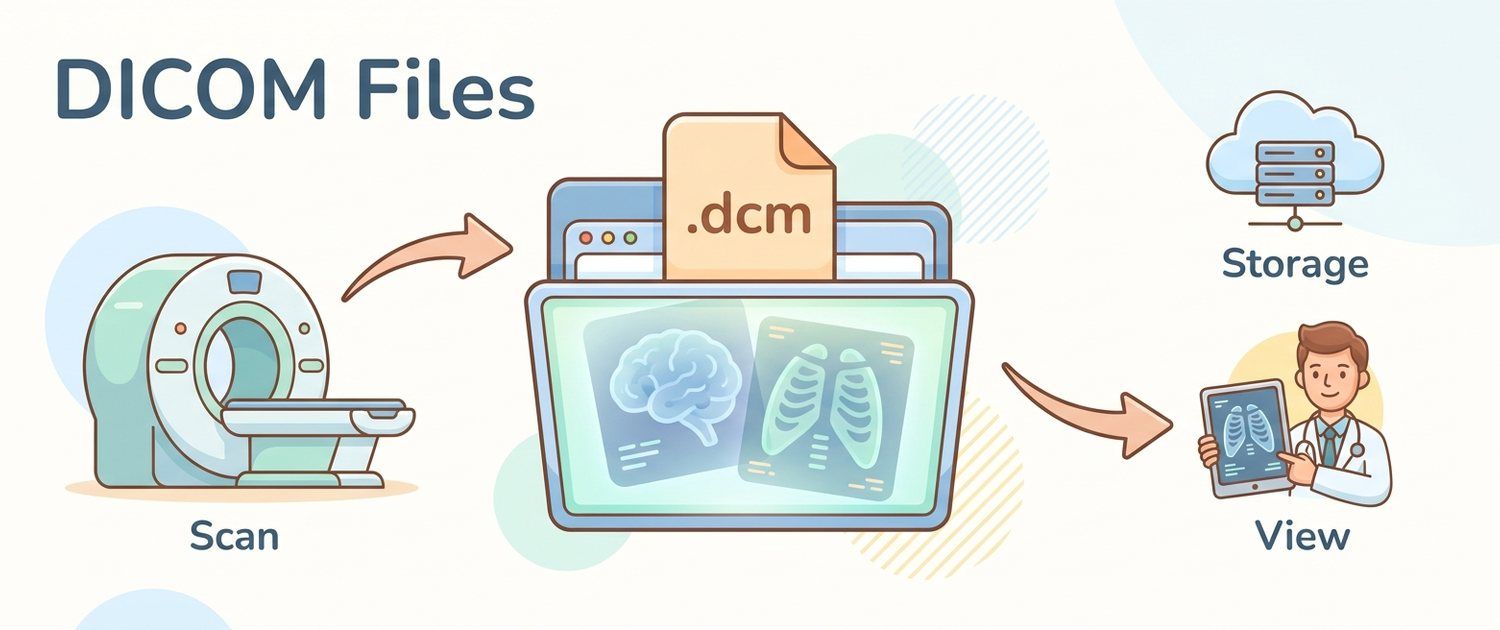
A .dcm file opens with neither an image viewer nor a browser, and sometimes it has no extension at all — just IM000001. It is a DICOM file, the format scanners use to store studies.
What DICOM is
DICOM is the international standard for storing and transmitting medical images. One .dcm file is usually one slice or one image; a full CT study can be hundreds of them in a single folder.
How it differs from JPEG
- Brightness scale. JPEG stores 256 shades of grey; DICOM usually 4096 or more. That is why a viewer can change the "window" and show lungs, then liver, then bone on the same slice — a JPEG keeps only the one view chosen at export.
- Units. On CT, brightness maps to physical tissue density in Hounsfield units: air about −1000, water 0, bone above 700. That is how a cyst is told from a solid mass. A JPEG keeps only dimensionless brightness.
- Acquisition data. The header records modality, projection, laterality, slice thickness, pixel size in millimetres and contrast use — none of which can be recovered from the picture.
So keep the originals: a screenshot is fine for showing someone, but part of the data is already gone from it.
Free viewers for Windows and macOS
All of these viewers are free to use. Windows: MicroDicom (free for personal use, portable build available), RadiAnt (free trial mode), Weasis (free and open). macOS: Horos, Weasis. Mobile options exist in both app stores, but fine detail — usually the part that decides the reading — shows poorly on a phone.
Open the whole folder, not a single file — then the slices assemble into a series you can scroll.
Common problems
- No extension — normal, the standard doesn't require one; the format is recognised by content.
- Only one slice opens — you opened a file instead of the folder.
DICOMDIRwon't open — it is the disc's table of contents, not an image. See how to open an MRI or CT disc.- "Format not supported" — the pixels inside may be compressed (JPEG 2000, JPEG-LS, RLE) and not every viewer knows them all; try Weasis or RadiAnt.
Online options and conversion
Online viewers exist and run in the browser with no program to install, but DICOM headers carry the patient's name and date of birth in plain text — check what such a service promises to do with your upload before choosing among these options. Conversion to JPEG is one-way: the brightness scale, densities and acquisition data are lost. Convert a copy, keep the originals.
If you need to understand, not just open
In imaging decoding .dcm files are uploaded as they are and read directly — no viewer needed. The original beats a photo of a screen: viewing windows are rebuilt from the full brightness scale, density is measured in Hounsfield units, and projection, laterality and contrast come from the header. This is informational support and does not replace a doctor.
For pet studies from a vet clinic disc, the same applies: pet imaging decoding.
Most of what comes on a vet clinic disc is CT and MRI — how the two differ in animals and what each of them shows is covered in MRI and CT for pets. A plain vet X-ray is often handed over as a printout or a photo of the film rather than a file; how such an image is read is covered in dog X-ray. Ultrasound is almost never saved as DICOM at all — you are left with single frames and the vet's description, and what echogenicity and free fluid mean there is unpacked in abdominal ultrasound for dogs.
See also: MRI interpretation, PET-CT, ECG interpretation.
For informational purposes only
This article is for informational purposes only and does not constitute medical advice, diagnosis, or treatment. Please consult a healthcare professional for medical guidance.