Dog X-Ray: What It Shows in the Chest, Abdomen and Joints
Reviewed by the LabReadAI medical team
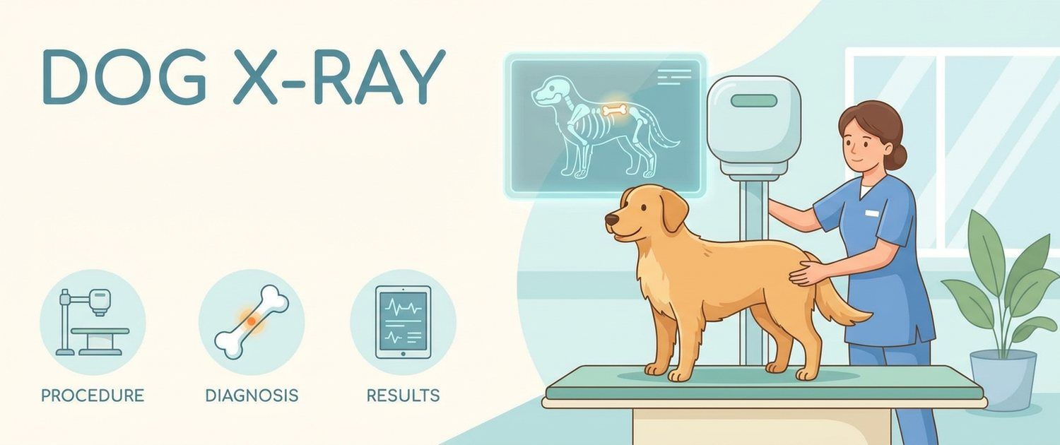
An X-ray is still the fastest way to look inside a dog: the image takes minutes, costs little and answers the questions that matter in the first hour — is there a fracture, free gas, a swallowed object, are the lungs flooded. Then the hard part starts: the owner looks at a grey-and-black picture and sees nothing at all. Let's go through what a dog X-ray shows, how to prepare for it and how to read the image together with the report.
What a Dog X-Ray Shows
An X-ray distinguishes only five densities, and that is both its strength and its ceiling:
| Density | How it looks | What it is |
|---|---|---|
| Gas | Black | Lungs, air in the bowel |
| Fat | Dark grey | Fat tissue, organ outlines |
| Soft tissue and fluid | Grey | Muscle, organs, blood, effusion |
| Bone | White | Skeleton, teeth, calcium stones |
| Metal | Bright white | A pellet, needle, pin, coin |
The key consequence: fluid and soft tissue are indistinguishable. Effusion in the chest therefore erases the outline of the heart, and a soft foreign object in the bowel is invisible. Where exactly those two densities must be told apart, ultrasound takes over — for example a dog abdominal ultrasound.
When a Dog Needs an X-Ray
- trauma: a fall, a road accident, a fight, a suspected fracture;
- limping and leg pain — what gets imaged then is covered in a limping dog;
- coughing, breathlessness, noisy breathing — see a coughing dog;
- a suspected foreign body: the dog swallowed a bone, a stone, a toy;
- repeated vomiting in dogs and a distended abdomen;
- routine lung screening in cancer patients;
- sizing the cardiac silhouette;
- assessing a young dog's joints before breeding clearance.
If you are still gathering the picture and the appointment is not today, it helps to describe your symptoms or work through the dog symptoms section, so the vet sees the trend rather than a single episode.
Dog Chest X-Ray: Lungs and Heart
The chest is imaged in at least two views — lateral and frontal — and in three when metastases are the question, because some nodules show on one side only. The radiologist describes the lung pattern as bronchial, alveolar or interstitial: these are ways diseased tissue looks, not separate diseases.
The heart is sized by its silhouette, most often with the vertebral heart score, which compares the heart's length to the thoracic vertebrae. It is a coarse ruler: it says whether the heart is enlarged, not which chamber or what the valves are doing. Those questions belong to echocardiography, covered in dog heart ultrasound and ECG.
Dog Abdomen X-Ray and Foreign Bodies
On an abdominal image the vet looks for gas where none should be, displaced organs, an enlarged liver or spleen, stones in the bladder and kidneys, and signs of obstruction.
An important limit: radiolucent objects — fabric, plastic, rubber, a sock — do not appear at all. Indirect signs give them away: dilated bowel loops, an abrupt change in calibre, trapped gas. That is why a suspected obstruction often means a repeat image hours later, or added ultrasound.
A separate emergency is gastric torsion in large breeds. It has a very characteristic look on a lateral image, and the clock matters: a distended abdomen, unproductive retching and restlessness mean going in immediately rather than waiting for morning.
Spine, Joints and Hip Dysplasia
Bone is the method's home ground. An image shows fractures, dislocations, arthritic new bone, changed joint shape and bone tumours. Hip dysplasia is assessed on a strictly extended positioning: without correct placement the grade is unreliable, so this study is nearly always done under sedation — a decision the vet makes. Orthopaedic images are covered in detail in X-ray of a dog's legs and joints.
The spine is the least rewarding region: a plain image shows vertebrae, narrowed spaces and a calcified disc, but not the disc material itself or the spinal cord. If a dog has weak hind legs or back pain, the image is only step one, and the next step is MRI and CT for dogs and cats.
How to Prepare a Dog for an X-Ray
- Abdomen — fasting for 8–12 hours unless it is an emergency: stomach content blocks the view.
- Collars and metal fittings come off: metal leaves a bright blot over what matters.
- Chest images need no preparation.
- Calm matters more than owners expect: the frame is taken on a breath-hold, and a dog that jerks earns a blurred image and a repeat.
- Some studies in animals are done under sedation — for orthopaedic positioning, marked pain or strong resistance. The vet decides.
How a Veterinary X-Ray Is Done and Whether It Is Safe
The dog is placed on the table, steadied with sandbags or by staff in protective gear, and the exposure lasts a fraction of a second. Owners are usually not allowed in the room — that is radiation safety, not mistrust.
The dose from one study is small and the diagnostic gain almost always outweighs it. "Almost always" is not "indiscriminately", though: pregnant animals and puppies are imaged on indication, and repeat series are taken when a decision depends on them.
How to Read a Dog X-Ray and Its Report
A radiologist's report is a description, not a diagnosis. Typical phrases and what they mean:
| Phrase | What it usually means |
|---|---|
| Increased lung markings | Non-specific; seen in healthy older dogs too |
| Alveolar pattern | Alveoli are filled — with fluid, pus or blood |
| Enlarged cardiac silhouette | Needs echo to name the chamber and the cause |
| Free gas in the abdomen | Almost always an emergency |
| Signs of obstruction | Urgent assessment and a decision are needed |
| Periosteal reaction | Bone has responded to a process; many causes |
| Loss of abdominal detail | Often fluid or little fat; clarified by ultrasound |
Looking at the image yourself is useful, with one caveat: ordinary screens and phones flatten the detail, so "I cannot see anything" means nothing. How findings on an image relate to blood numbers is shown by pet results interpretation, and canine reference ranges are covered in the dog blood test article.
X-Ray Versus Ultrasound, CT and MRI
- X-ray — fast, cheap, available; bone, lungs, gas, metal.
- Ultrasound — soft tissue, fluid, organ walls, motion; no radiation.
- CT — the same physics, but in slices and in volume; the best method for lungs and complex fractures.
- MRI — soft tissue, brain and spinal cord, intervertebral discs.
The usual order is: image first, next step by result. Rabbits, rodents and birds have their own positioning and approach, gathered in imaging for small pets. Cats shift the emphasis — what is looked at in them is covered in cat X-ray.
When an Image Calls for Urgent Action
Do not wait if the description mentions free gas in the abdomen, signs of gastric torsion, a large chest effusion, pneumothorax, a displaced fracture or a suspected ruptured organ. With severe breathlessness the study is sometimes postponed until the animal is stable — that is sound practice, not a refusal of care.
If the image is already in your hands, upload it (a photo of the film, a PDF or files from the disc) to a pet scan interpretation: the description is explained in plain language for the species and turned into questions for your vet. If the disc will not open, see how to open an MRI or CT disc and how to open a DICOM (.dcm) file.
This article is informational. Diagnosis and treatment of your dog are the job of a veterinarian.
For informational purposes only
This article is for informational purposes only and does not constitute medical advice, diagnosis, or treatment. Please consult a healthcare professional for medical guidance.