Dog Abdominal Ultrasound: What It Shows and How to Read It
Reviewed by the LabReadAI medical team
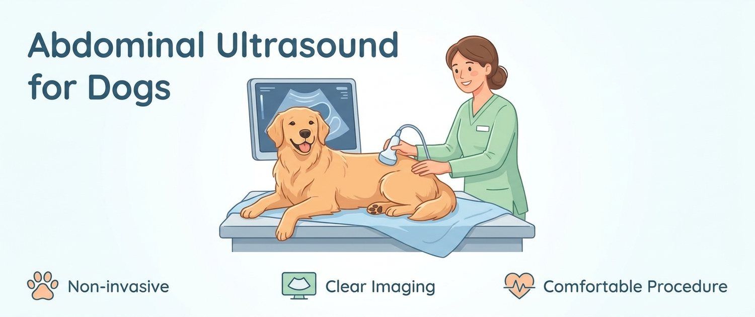
An abdominal ultrasound is the study a dog is referred for most often when someone needs to see what is happening inside. It needs no incision, uses no radiation and shows organs moving in real time. The difficulty comes afterwards: the owner receives a page full of phrases like "parenchymal echogenicity is increased" and cannot tell whether the document is alarming or calm. Let's go through what a dog abdominal ultrasound shows, how to prepare the animal and how to read the report.
What a Dog Abdominal Ultrasound Shows
The vet moves the probe across the belly and works through the liver and gallbladder, spleen, kidneys and bladder, adrenal glands, stomach and loops of intestines, pancreas, abdominal lymph nodes, and in intact animals the prostate or the uterus and ovaries.
The key idea about the method: an abdominal ultrasound shows structure — organ size, shape, tissue uniformity, stones, cysts, masses, free fluid. It does not show how an organ works. Function is assessed by blood, so the report and the dog blood test are read together: a kidney may look almost ordinary while kidney markers are already rising, and the reverse happens too.
It is just as important to know what ultrasound cannot do. It sees poorly through gas and bone: lungs, bowel content, the inside of the skull. A cough or an injury therefore calls not for an abdominal scan but for a dog X-ray, while difficult head and spine cases go to MRI and CT for dogs and cats.
When a Dog Needs an Ultrasound
Typical reasons an owner walks out of the consulting room with a referral:
- repeated vomiting in dogs, especially when it does not settle within a day;
- prolonged diarrhoea in dogs, or diarrhoea alternating with constipation;
- a dog not eating, losing weight or turning lethargic for no clear reason;
- changes in bloodwork — rising liver or kidney markers, unexplained anaemia;
- suspected foreign body and obstruction: the dog swallowed a toy, a bone, a sock;
- frequent or painful urination, blood in urine, suspected stones;
- a swollen abdomen, pain on palpation, the "praying" posture;
- pregnancy monitoring and routine screening in an older animal.
When the signs are there but the referral is not yet, it helps to gather the picture first: you can describe your symptoms or start from the dog symptoms section, so the vet sees a trend rather than a single day.
How to Prepare a Dog: Fasting, Fur and Calm
Preparation affects the result more than owners expect. Gas and food scatter ultrasound, and the pancreas and adrenal glands simply drop out of view.
| What is done | Why |
|---|---|
| Fasting for 8–12 hours | Removes food and gas that block the view |
| Water is not withheld | Dehydration worsens both picture and patient |
| A full bladder | The bladder wall and stones are visible only against urine |
| Clipped fur on the belly | Without probe-to-skin contact there is no image |
| A calm setting | A tense abdominal wall obstructs the scan as much as gas |
In puppies and toy breeds the fasting window is shorter — the clinic gives the exact time. Some studies in animals are done under sedation: that decision belongs to the vet, based on the dog's condition and temperament, and the owner should give nothing on their own.
How a Veterinary Ultrasound Is Done
The dog lies on its back or side in a padded trough, gel is applied and an assistant steadies the animal. The scan takes 20–40 minutes — longer than a human study, because there are many organs and each is checked in two planes. It is painless; the worst of it is cool gel and the need to lie still.
If the vet sees a suspicious area, a sample can be taken there and then under probe guidance — a fine-needle aspirate. That is a separate procedure and you are warned about it in advance.
How to Read a Dog Ultrasound Report
The report is written descriptively: the vet records what is visible rather than naming a disease. That is why the page feels full of threats when most of the wording is neutral.
| Phrase | What usually lies behind it |
|---|---|
| Smooth contours, homogeneous structure | Normal |
| Echogenicity increased / decreased | Tissue brighter or darker than usual — non-specific |
| Diffuse parenchymal changes | Spread through the organ, not a focus; cause to be found |
| Calculi, shadowing hyperechoic foci | Stones or dense inclusions |
| Sediment in the bladder | Debris, common outside disease as well |
| Thickened bowel wall, loss of layering | Inflammation or infiltration — to be clarified |
| Free fluid | The cause must always be established |
| Mass, nodule | Further work-up needed, often a needle sample |
One reading rule matters: a description is not a verdict. Increased liver echogenicity occurs in a healthy older dog and in serious disease alike; what separates them is the combination with signs and biochemistry, not the phrase itself.
Liver, Kidneys and Bladder: Increased Echogenicity and Other Phrases
For the liver the vet judges size, contours, uniformity and the gallbladder. An isolated note about echogenicity with unchanged bloodwork usually means monitoring rather than treatment.
For the kidneys it is size, the corticomedullary boundary, pelvic dilation and stones. For the bladder it is wall thickness, sediment and calculi — this is where the reason for frequent, painful urination is most often found.
The spleen alarms owners more than any other organ: nodules are common in older dogs and far from all of them are dangerous. Working through such a finding alone online is pointless — it needs a vet's assessment. The general logic of veterinary sonography is covered in ultrasound for dogs and cats, and the feline specifics in cat abdominal ultrasound.
Free Fluid and Masses: What They Mean
Free fluid in a dog's abdomen is the most common worrying finding. On its own it is not a diagnosis: fluid appears after trauma, in liver and heart disease, in inflammation, and with bleeding from a mass. What matters is how much there is, how the animal looks and what the blood says. A small volume in a bright dog and a growing volume in a dog with pale gums are two different situations.
The word "mass" does not equal cancer either: cysts, regenerative nodules, haematomas, abscesses and tumours — benign and malignant — all hide behind it. They cannot be told apart by the picture, which is exactly why sampling exists.
Ultrasound Versus X-Ray, CT and Blood Tests
- Ultrasound — soft tissue, fluid, motion; safe and repeatable.
- X-ray — bones, lungs, gas, foreign bodies, the whole abdomen in one frame.
- CT and MRI — volume and precise anatomy where the other methods run out.
- Blood and urine — organ function; what those numbers mean is covered by pet results interpretation.
- The heart is a separate story, examined by echocardiography — see dog heart ultrasound and ECG.
The methods do not replace each other. If a dog is limping, an abdominal scan will not help — a joint image will; and a leg image is useless in a vomiting dog.
When Findings Mean Going to the Vet Urgently
Do not delay if the report shows free fluid together with lethargy and pale mucous membranes, signs of obstruction or torsion, a large mass with breakdown, a ruptured organ, or a rapidly enlarging kidney with a dilated pelvis. Urgency is set less by the wording than by the dog: deterioration over hours always means going in, even with "moderate" phrasing in the report.
If the report is already in your hands and you want every line in plain language, upload it (a PDF, a photo of the page or frames from the study) to the veterinary imaging review: the service explains the wording for the species and prepares questions for your vet. If the clinic handed you a disc and the files will not open, see how to open an MRI or CT disc and how to open a DICOM (.dcm) file — you can upload them as they are.
This article is informational. Diagnosis and treatment of your dog are the job of a veterinarian.
For informational purposes only
This article is for informational purposes only and does not constitute medical advice, diagnosis, or treatment. Please consult a healthcare professional for medical guidance.