Cat Abdominal Ultrasound: What It Shows and How to Read It
Reviewed by the LabReadAI medical team
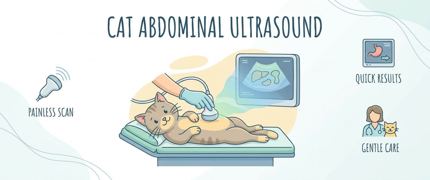
A cat rarely gives a clear signal: she does not limp for effect and does not complain, she simply stops jumping onto the windowsill and sleeps more. That is why ultrasound is often the first method that turns a vague "something is off" into concrete lines on a page. Let's go through what a cat abdominal ultrasound shows, how to prepare the animal and how to read the report without panicking at descriptive words.
What a Cat Abdominal Ultrasound Shows
In one session the vet works through the liver and gallbladder, spleen, both kidneys, bladder, adrenal glands, stomach and intestines, pancreas, lymph nodes, and in intact females the uterus and ovaries.
Ultrasound shows structure: size, contours, tissue uniformity, wall thickness, stones, cysts, nodules and free fluid. It does not measure how an organ works — blood does that, so the report and the cat blood test only make sense together. The classic example: kidneys already look small and irregular on the image, while blood shows how far function has actually slipped.
What a veterinary ultrasound will not show is lungs, bones and anything hidden behind gas. Those belong to a cat X-ray, and complex head or spine anatomy goes to MRI and CT for dogs and cats.
When a Cat Needs an Ultrasound
- weight loss with a preserved or even increased appetite;
- vomiting and diarrhoea in cats, especially repeating over weeks;
- a situation where why a cat is not eating stays unanswered for more than a day;
- thirst and larger urine volumes — the classic reason to look at kidneys;
- blood in a cat's urine, frequent litter tray visits, crying while urinating;
- suspected foreign body: cats swallow thread, tinsel, hair ties;
- a firm lump the owner has felt themselves;
- monitoring an older animal or a known chronic disease.
If the signs are there but the appointment is a day or two away, gather observations first: you can describe your symptoms or start from the cat symptoms section, so the vet sees a trend rather than one episode.
How to Prepare a Cat: Fasting, Fur and Calm
| What is done | Why |
|---|---|
| 8–12 hours without food | Food and gas block the view |
| Water stays available | A dehydrated cat scans worse and copes worse |
| A full bladder | Wall and stones show only against urine |
| Clipped fur on the belly | No air may sit between probe and skin |
| Carrier and quiet | A frightened cat tenses the abdomen and quality drops |
Kittens get a shorter fasting window. Some studies in animals are done under sedation — the vet decides, weighing the cat's temperament and condition; the owner should give nothing independently.
How a Veterinary Ultrasound Is Done in Cats
The cat is placed on her back or side, warm gel is applied and an assistant holds her gently. The scan takes 20–40 minutes: feline organs are small, the probe is high-frequency, and each kidney is checked in several planes. The procedure causes no pain.
If something looks suspicious, the vet may take a fine-needle sample under ultrasound guidance right away — a separate procedure you are told about in advance.
How to Read a Cat Ultrasound Report
The report describes what was seen; it does not name a disease. Hence the abundance of words that sound alarming but mean "keep watching".
| Phrase | What usually lies behind it |
|---|---|
| Smooth contours, homogeneous structure | Normal |
| Increased cortical echogenicity | Common in older cats; read with bloodwork |
| Reduced corticomedullary distinction | The boundary between layers is blurred |
| Calculi in the bladder | Stones; the plan is the vet's |
| Sediment in the bladder | Debris, seen outside disease too |
| Thickened bowel wall | Inflammation or infiltration — to be clarified |
| Enlarged mesenteric lymph nodes | A reaction; the cause is sought separately |
| Free fluid | The cause must always be established |
The main reading rule: a description is not a verdict. The same phrase means different things in a young and an elderly cat, and it is the combination with signs and tests that decides, not the word.
Kidneys and Bladder: Why Cats Get Them Checked First
The urinary system is this species' weak spot. For kidneys the vet judges size, contour smoothness, cortical thickness, pelvic dilation and stones. Small irregular kidneys in an elderly cat and one enlarged kidney with a dilated pelvis are entirely different stories, and the second is often urgent.
For the bladder it is wall thickness, sediment and calculi. This is where the explanation for frequent tray visits and crying is usually found; what changes about feeding once stones are confirmed is covered in feeding a cat with urinary stones.
Intestines, Liver and Pancreas
In cats the intestine is the second most common source of findings. The vet measures wall thickness and judges its layering: even thickening with preserved layers and focal thickening with lost layers are read differently, and the second nearly always leads to further work-up.
The liver is judged by size, contours and uniformity; an isolated note on echogenicity with unchanged biochemistry usually means monitoring. The pancreas is not always visible in cats — its absence from the report does not mean something is wrong with it. The general logic of the method in both species is covered in ultrasound for dogs and cats, and the canine specifics in dog abdominal ultrasound.
Free Fluid and Masses in Cats
Free fluid in a cat's abdomen always needs explaining. It follows trauma, liver and heart disease, inflammation, bleeding and infectious processes. Volume, the fluid's character on sampling and the animal's state all matter: lethargy, pale gums and fast breathing turn a finding into an emergency.
"Mass" in a report is not a synonym for cancer either: cysts, nodules, haematomas, abscesses and tumours all sit behind the word. A single image cannot tell them apart, so sampling usually comes next.
Ultrasound Versus X-Ray and Blood Tests
- Ultrasound — soft tissue, fluid, organ walls, motion; no radiation and repeatable.
- X-ray — bones, lungs, gas and the whole abdomen in one frame.
- Blood and urine — function; what the numbers mean is explained by pet results interpretation.
- The heart is examined separately by echocardiography — the principle is covered in dog heart ultrasound and ECG and holds for cats too.
- Rabbits, rodents and birds follow their own rules, gathered in imaging for small pets.
To go through the report line by line, upload it (a PDF, a photo of the page or frames from the study) to the pet imaging decoding service: the wording is explained for the species and you get questions to ask your vet. If the clinic gave you a disc that will not open, see how to open an MRI or CT disc.
This article is informational. Diagnosis and treatment of your cat are the job of a veterinarian.
For informational purposes only
This article is for informational purposes only and does not constitute medical advice, diagnosis, or treatment. Please consult a healthcare professional for medical guidance.