X-Ray and Ultrasound for Rabbits, Hamsters, Guinea Pigs, Birds
Reviewed by the LabReadAI medical team
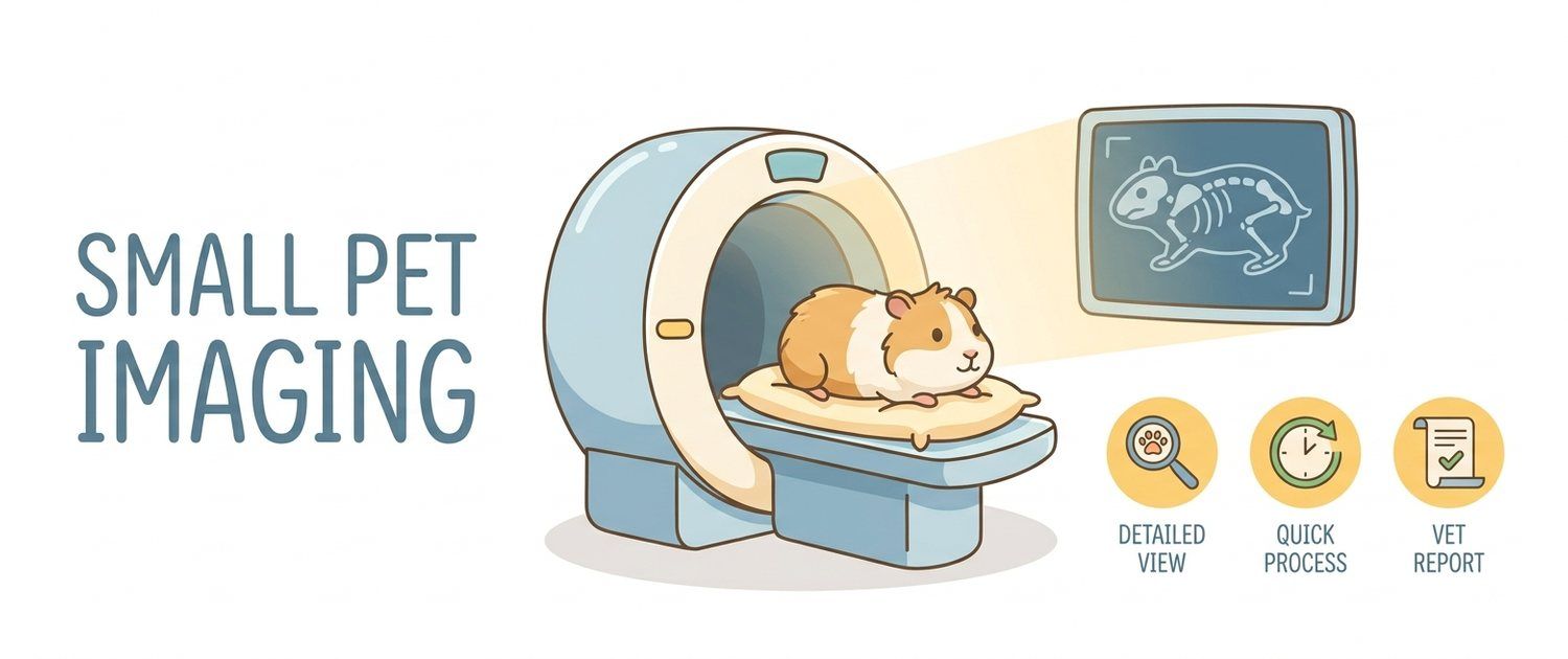
With a dog or a cat the order is familiar: examination, blood work, then a picture. With a small pet that order breaks. In a 40-gram hamster a full biochemistry panel is barely possible; in guinea pigs and parrots drawing blood is itself a stress and a risk. So here imaging is often the first objective study of any kind — where the work-up starts rather than where it ends.
Why a Small Pet Gets Imaging Before Blood Tests
Three reasons the picture comes first:
- Blood volume. In an animal of a few dozen grams only a tiny sample can be taken safely, which leaves the laboratory very little room.
- Concealment. Rabbits and rodents are prey species, and evolution taught them to hide illness. Owners notice a problem once compensation has already failed.
- Reference ranges. Norms for many small species are known far less well than for dogs and cats. How a report is read where norms are solid is shown by pet results interpretation.
A film has none of those limits: it works the same in a rabbit and in a rat, and it takes seconds.
What a Rabbit X-Ray Shows
Four regions are read in rabbits:
- Skull and teeth — the most common and most important;
- Abdomen — gas distension of the stomach, stagnant content, signs of gut stasis;
- Chest — heart, lungs, mediastinal masses;
- Urinary tract — sludge and stones, frequent in this species.
Gut stasis — the state in which a rabbit stops eating and passing droppings — appears as a gas-distended stomach with immobile content. Telling simple stasis from true obstruction is only possible on the film, and that decision changes the whole plan.
Rabbit and Rodent Teeth: the Main Reason for a Film
In rabbits, guinea pigs and chinchillas every tooth grows continuously; in rats and hamsters only the incisors do. When wear goes wrong, crowns drift sideways and roots travel deep into the jaw. The owner sees drooling, refusal of hard food and weight loss, while the cause is buried in bone.
The film is read for root length and direction, jaw bone quality, abscesses and orbital involvement. An oral exam shows only the tip of the problem — literally. That is why dental imaging is the standard in rabbits and rodents, and complex cases add cross-sectional imaging, the principle behind MRI and CT in dogs and cats.
Hamster, Rat and Chinchilla X-Rays
In hamsters a film is usually taken after trauma (a fall, a trapped limb), for a suspected tumour or for abdominal distension. Rats have their own big theme — lungs: chronic respiratory infection is near-inevitable, and radiography shows how far it has gone. In chinchillas, as in rabbits, teeth come first.
All small species share one difficulty: size. The resolution of a standard veterinary unit is already at its limit on a 30–100 gram patient, so clinics use dedicated settings and magnified positioning.
Guinea Pig X-Ray and Bladder Stones
In guinea pigs stones in the bladder and ureters are among the commonest findings, and nearly all of them show well because they contain calcium. Clinically this looks like straining, discomfort on urination and blood in the urine.
The second recurring theme is again teeth, the third is lungs. In females the ovaries are assessed too: cysts are regular in guinea pigs and show better on ultrasound.
Parrot and Bird X-Ray
Bird films read differently: birds have air sacs and hollow bones, and the liver, heart and stomach form a characteristic silhouette. What is usually assessed:
- bone density — it changes in laying females, and that is normal;
- egg binding — a calcified egg shows clearly;
- crop and stomach — foreign bodies, metal, grit;
- air sacs — opacity points to inflammation;
- liver — an enlarged silhouette is a frequent finding in parrots.
Metal in a parrot's stomach is a reason of its own to take a film: birds chew hardware and coatings, and radiography finds such fragments at once.
Ultrasound in Rabbits and Small Rodents
Ultrasound is used less often than radiography in small species, but within its zone it is irreplaceable: heart, uterus and ovaries, kidneys, bladder, free abdominal fluid. In rabbits echocardiography helps with breathlessness, in guinea pigs with suspected ovarian cysts.
The limit is simple: bowel gas, and herbivorous rodents have plenty. The general logic is the same as in larger pets, described in ultrasound for dogs and cats; the same territory in cats is covered by cat abdominal ultrasound.
Anaesthesia and Handling: What Small Species Risk
A small animal loses heat fast, reacts to stress faster and tolerates prolonged restraint badly. Hence the rule: the study must be short.
Many films are taken with no sedation at all — in a clear chamber or with gentle restraint, in seconds. Sedation is added when strict positioning is needed (skull, teeth) or when struggling makes injury more likely than the drug does. The vet decides after assessing the patient; in dogs and cats the same fork looks different — see dog leg and joint X-ray. The basics of plain film are in dog X-ray, and the large-animal version is in horse X-ray and ultrasound.
Reading the Report
| Phrase | What it usually means |
|---|---|
| Elongated tooth roots, jaw deformity | The classic dental disease picture in rabbits and rodents |
| Gas distension of the stomach | A sign of stasis or obstruction, needs context |
| Radiopaque calculi in the bladder | Stones, a common finding in guinea pigs |
| Reduced lung field aeration | Possible inflammatory process |
| Air sac opacity (in birds) | A sign of respiratory inflammation |
| Metallic densities in the stomach | Swallowed metal, frequent in parrots |
If the clinic handed you a disc or files, see how to open an MRI or CT disc and how to open a DICOM (.dcm) file. Upload the image or a photo of the report to pet imaging interpretation — the service explains the wording for the species. If there is no film yet, you can describe the symptoms and see where to begin.
This article is informational. Diagnosis and treatment of your pet are the job of a veterinarian.
For informational purposes only
This article is for informational purposes only and does not constitute medical advice, diagnosis, or treatment. Please consult a healthcare professional for medical guidance.