Tortoise and Reptile X-Ray: Why It Is Done and What It Shows
Reviewed by the LabReadAI medical team
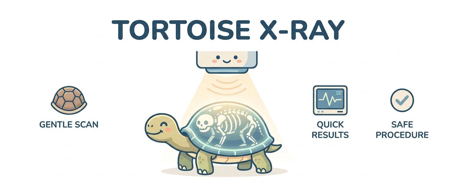
With a dog you can see that it limps or breathes hard. With a tortoise you see almost nothing: it is barely mobile when healthy and barely mobile when dying. That is why a tortoise x-ray is not an extra test but often the first way to learn anything at all about what is happening inside.
Why a Tortoise or Reptile X-Ray Is Done
The reasons owners come to the clinic:
- the tortoise stopped eating, went quiet, will not leave its hide;
- a female is restless and digging but lays no eggs;
- the shell feels soft, the legs splay, the beak is deforming;
- the animal wheezes, breathes with an open mouth, blows bubbles from the nose;
- it swallowed a pebble, substrate or a piece of decor;
- there was trauma — a fall, a car, a dog bite.
A film answers nearly all of these quickly and cheaply. In mammals ultrasound plays that role, but in reptiles access is limited — compare ultrasound for dogs and cats. The same logic holds for rodents and birds, covered in detail in small pet imaging.
Metabolic Bone Disease: the Main Finding
Metabolic bone disease is the commonest thing found in pet reptiles, and it is nearly always a consequence of husbandry: too little UVB light, an unbalanced calcium-to-phosphorus ratio in the diet, temperatures too low for metabolism to run at all.
On the film it appears as reduced bone density: the cortex is thin, the bones look translucent, the shell loses its crisp outline, and pathological fractures and jaw deformity turn up. The vet compares bone density with shell density — in advanced disease the difference disappears.
A film shows the state, not the treatment. Correcting lighting, temperature and diet is the vet's job and depends on species and age; blood work is often run alongside, and pet results interpretation explains what those numbers mean.
Egg Retention in a Tortoise: When the Film Decides
Tortoise eggs are calcified, so they show beautifully on an x-ray — they can be counted, measured and placed. This is where the film is irreplaceable: a restless female before a normal clutch and a female with retained eggs look identical from outside.
The vet checks how many eggs there are, whether they are deformed or too large for the pelvis, and how long the process has been running. Everything that follows — from a proper nesting site to surgery — depends on that. Trying to force laying at home is dangerous.
Foreign Bodies and Obstruction
Tortoises and lizards regularly swallow substrate, stones, sand, silicone and fishing line. Dense objects show at once, and their position tells whether they are stuck or moving. Some of these finds are not accidents: an animal short of minerals starts eating its own substrate.
Radiolucent objects — soft plastic, fabric — do not show directly and are judged indirectly, from trapped gas and dilated bowel loops.
Pneumonia and Airway Disease in Reptiles
Reptiles have no diaphragm and simpler lungs than mammals: inflammatory exudate is not coughed up but pools in place. Pneumonia in a tortoise is therefore severe and often drawn out.
The film is read for shadowing of the lung field, asymmetry between the two lungs and fluid levels. In chelonians this requires lateral and craniocaudal views: on a dorsoventral film the lungs are completely covered by the shell.
The Shell as an Obstacle and a Landmark
The shell is dense bone, and it wipes out the detail of everything beneath it. Two practical consequences follow.
First, soft tissue in a tortoise is far less legible than in a dog or a cat, so a single view is almost never enough — three views are the standard. Second, the shell itself is a good indicator: its thickness, density, outline and symmetry tell the story of mineralisation and of old injuries. What the shell hides on a plain film is sometimes recovered with cross-sectional imaging, the principle behind MRI and CT in dogs and cats.
What a Lizard or Snake X-Ray Shows
In lizards the same things are read: bone density, fractures, eggs, foreign bodies, lungs. In snakes a film helps locate a swallowed object, assess the long and easily injured spine, and show eggs or embryos.
Reptiles rarely need anaesthesia for radiography: they are placed in a restraint chamber or on a stand and their cold-blooded metabolism lets them lie still. That sets them apart from dogs and cats, which often need sedation for strict positioning — see dog leg and joint X-ray.
Reading the Report and What Comes Next
| Phrase | What it usually means |
|---|---|
| Reduced bone mineral density | The metabolic bone disease picture |
| Thinned cortical bone | Bone is losing strength, fracture risk |
| Calcified eggs in the pelvic area | A clutch has formed; the question is whether it is moving |
| Radiopaque foreign body | A dense object in the gut, position assessed |
| Lung field opacity | Possible pneumonia, needs clinical context |
| Shell deformity, pyramiding | The mark of long-standing husbandry problems |
Clinics may hand out a disc or DICOM files. If they will not open, see how to open an MRI or CT disc and how to open a DICOM (.dcm) file.
Upload the image or a photo of the report to pet imaging interpretation — the service explains the wording for the species and builds the questions to ask a reptile vet. If there is no film yet, you can describe the symptoms and see where to start. Large-animal imaging follows the same logic at a different scale, described in horse X-ray and ultrasound, while the basics of plain film are in dog X-ray.
This article is informational. Diagnosis and treatment of your pet are the job of a veterinarian.
For informational purposes only
This article is for informational purposes only and does not constitute medical advice, diagnosis, or treatment. Please consult a healthcare professional for medical guidance.