Horse X-Ray and Ultrasound: What Vets Look For in the Images
Reviewed by the LabReadAI medical team
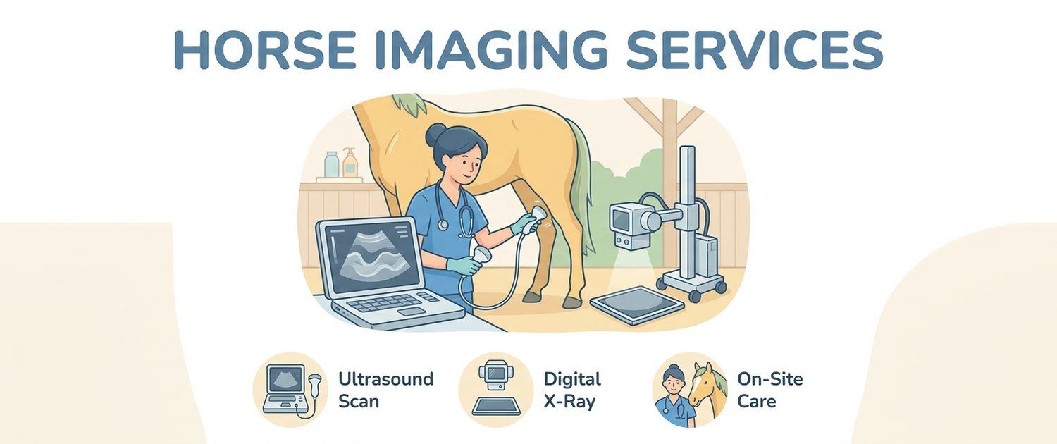
A horse weighs half a tonne and does not fit into a scanner. So equine imaging is organised the other way round from small animals: the machine travels to the patient, and almost all the work rests on two methods, radiography and ultrasound. Here is what vets look for with each and how to read the report you are handed.
What Vets Look For on a Horse X-Ray
Portable radiography in horses is mainly an orthopaedic tool. Limbs are imaged most, then the head, neck and back. The vet assesses:
- bone outline and density, fractures and fissures;
- joint spaces and marginal bony outgrowths;
- the position of the coffin bone inside the hoof capsule;
- teeth and sinuses when there is facial swelling or one-sided discharge;
- the spinous processes of the vertebrae in suspected back problems.
The chest of an adult horse cannot be penetrated properly by a standard portable unit, so lungs are usually assessed by ultrasound and airway endoscopy instead. That is a fundamental difference from small animals, where a chest film is routine — see dog X-ray.
Hoof X-Rays: the Main Site of Lameness
More than half of equine lameness lives inside the hoof, and the film settles most of it. The vet reads the position of the coffin bone relative to the hoof wall and sole, the navicular bone, signs of laminar inflammation, foreign bodies and nails, and changes after shoeing.
In laminitis the film shows rotation and sinking of the coffin bone — something invisible from outside until it is late. That is why hoof films are also taken electively: they let the farrier and the vet work from real anatomy rather than by eye.
Joint and Back X-Rays
Joint films — fetlock, hock, carpus, stifle — are taken for lameness, swelling and as part of a pre-purchase examination. The search is for osteophytes, subchondral bone cysts, signs of osteochondrosis and fragments inside the joint.
The equine back is a difficult region: the spacing of the spinous processes, their contact (kissing spines) and the facet joints are assessed. The logic of reading bone images is the same in a horse and in a dog, only the scale differs — it is set out in dog leg and joint X-ray.
What a Horse Ultrasound Shows
Ultrasound carries more weight in horses than in small animals, because it takes over part of the job cross-sectional imaging does there. The main areas:
- tendons and ligaments of the limbs — the principal field;
- abdomen in colic;
- chest — pleural fluid and the lung surface;
- heart — murmurs, arrhythmias, poor performance;
- reproductive tract — cycle monitoring and pregnancy.
The principle is the same as in small animals, described in ultrasound for dogs and cats; the cardiac part is covered in dog heart ultrasound and ECG.
Tendon and Ligament Ultrasound
This is what ultrasound exists for in the horse world. The superficial and deep digital flexor tendons and the suspensory ligament account for a huge share of all lost work in sport horses.
The scan is read for cross-sectional area, echogenicity, fibre alignment and any hypoechoic zone — the damaged part. Its key strength is follow-up: repeating the study over weeks and months shows whether healing is on track. That is why reports carry areas and percentages — they are a baseline for the next scan.
Abdominal Ultrasound and Colic
Colic is the commonest equine emergency, and ultrasound works here as a rapid triage tool. Within minutes the vet checks standard windows along the flank and belly and answers the key questions: are there distended small intestinal loops, are the walls thickened, is there free fluid, where do the stomach and spleen sit.
Those answers decide the main thing — surgery now or watchful waiting. No laboratory test works that fast.
How Imaging Is Done in the Stable
The usual order:
- the horse is stood on level, firm ground and the feet are picked out;
- for hoof films the foot is set on a wooden block to catch the right angle;
- for tendon scans the hair over the cannon is clipped, otherwise the image is murky;
- the horse stays on its feet; light sedation is used if it will not settle, chosen by the vet;
- everyone nearby wears protection against scattered radiation.
That is a real advantage over dogs, cats and especially small pet imaging, where strict positioning usually calls for restraint or anaesthesia. Reptiles follow their own logic — see tortoise X-ray.
Blood Tests and Images Together
Images show structure, blood shows how the body is working. In simple lameness blood is rarely needed, but in colic, fever, weight loss and poor performance it is almost always taken: inflammation, dehydration, muscle enzymes and metabolic markers change how the picture is read. What the lines in the report mean is covered in horse blood test and pet results interpretation.
Reading the Report
| Phrase | What it usually means |
|---|---|
| Coffin bone rotation | The bone has rotated — the laminitis picture |
| Marginal osteophytes | Bony outgrowths, a sign of a long-standing process |
| Closely apposed spinous processes | Kissing spines, a frequent back finding |
| Hypoechoic zone within the tendon | The damaged area; size and percentage matter |
| Increased cross-sectional area | The tendon is swollen or remodelled |
| Distended small intestinal loops | A worrying sign in colic |
| Free peritoneal fluid | Needs an urgent explanation |
If the clinic handed over files or a disc that will not open, see how to open an MRI or CT disc and how to open a DICOM (.dcm) file. Cross-sectional imaging of the lower limb is available in some referral centres, and how those methods work is explained in MRI and CT in dogs and cats.
Upload the image or a photo of the report to pet imaging interpretation: the service explains the wording in plain language and prepares questions for the vet. If there is no study yet and the horse has simply changed its movement or appetite, you can describe the symptoms — the same route dog owners take with lameness, covered in dog limping.
This article is informational. Diagnosis and treatment of the animal are the job of a veterinarian.
For informational purposes only
This article is for informational purposes only and does not constitute medical advice, diagnosis, or treatment. Please consult a healthcare professional for medical guidance.