Dog Heart Ultrasound and ECG: What They Show and When to Do It
Reviewed by the LabReadAI medical team
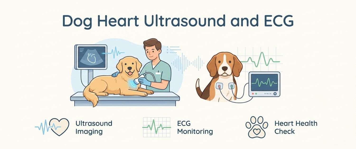
A dog's heart rarely hurts in a way anyone can see. The first sign is usually domestic rather than dramatic: the dog starts stopping on walks, coughs at night, breathes heavily after the stairs. Then the vet hears a murmur through the stethoscope and writes a referral for two studies at once — a heart ultrasound and an ECG. They do not duplicate each other: one looks at how the heart is built, the other at its electrics.
When a Dog Needs a Heart Ultrasound or ECG
- a murmur heard at a routine visit or before vaccination;
- coughing, especially at night and after exercise — why that is not always the lungs is covered in a coughing dog;
- breathlessness, fast breathing at rest, a bluish tongue;
- fainting or sudden weakness on a walk;
- poor exercise tolerance: an hour of running last year, ten minutes now;
- preparation for anaesthesia in an older animal or an at-risk breed;
- monitoring of known heart disease;
- an irregular pulse or an arrhythmia heard by the vet.
If you are still only noticing oddities, write them down first — describe your symptoms or work through the dog symptoms section, counting the breathing rate during sleep: it is one of the few home numbers cardiologists genuinely use.
What a Dog Heart Ultrasound Shows
Echocardiography is ultrasound aimed at the heart. The vet sees chambers, walls, valves and blood flow, and measures them:
| What is assessed | What it means |
|---|---|
| Left atrium and ventricle size | How volume-overloaded the heart is |
| Wall thickness | Thickening is the key sign of feline cardiomyopathy |
| Valve leaflets | Thickening and prolapse — the classic small-breed picture |
| Regurgitation (backflow) | Some blood returns; the volume is judged |
| Contractility | How forcefully the heart ejects blood |
| Pulmonary artery pressure | Indirect sign of pulmonary hypertension |
| Pericardial fluid | Needs its own, often urgent, plan |
Small breeds most often show age-related mitral valve degeneration, large breeds dilated cardiomyopathy. Both are diagnosed on echo: neither blood tests nor radiographs replace that picture. Blood is still needed — it shows how the heart affects kidneys and metabolism, and the numbers are explained in the dog blood test article.
How an ECG Differs From Echocardiography
An ECG shows neither size nor valves. It records the electrical signal and therefore answers a different question: does the heart fire and conduct correctly?
- Echo — anatomy and pump performance. Needed for murmurs, breathlessness, coughing, before anaesthesia.
- ECG — rhythm and conduction. Needed for fainting, an irregular pulse, poisoning, potassium shifts, before long procedures.
They complement each other: a fainting dog may have a perfectly normal echo and a dangerous arrhythmia, or the reverse. Ultrasound as a method is covered in ultrasound for dogs and cats, and the abdominal part of the exam in dog abdominal ultrasound.
Preparing the Dog and How the Exam Goes
No special preparation is needed: fasting is not required for echo and water is not withheld. Fur on both sides of the chest is usually clipped — without probe-to-skin contact there is no image. For the ECG, clips or adhesive electrodes are placed on the limbs and chest; this does not hurt.
The dog lies on its side on a table with a cut-out, and the study takes 20–40 minutes. Some studies in animals are done under sedation — the vet decides, weighing the animal's condition and temperament. With severe breathlessness the order reverses: stabilisation first, a full echo afterwards.
What the Heart Ultrasound Report Says
An echo report is a table of measurements plus a descriptive part. Three things usually alarm owners:
- Indexed values such as LA/Ao and LVIDDN. These are ratios scaled to body size, so a chihuahua and a mastiff can be compared. Crossing a threshold means chamber overload, not a countdown.
- Regurgitation. A small backflow occurs in healthy animals too; volume and whether the atrium has stretched are what count.
- Disease stages written as letters and numbers. That is clinicians' shorthand for whether structural change and symptoms are already present. Always ask the cardiologist what the stage in your report means — monitoring plans hinge on that letter.
How a Dog ECG Trace Is Read
An ECG trace is an image: millimetre paper with waves on it. That is exactly why it can be uploaded to a pet imaging interpretation alongside a radiograph or an ultrasound report — the curve itself is read, not only the text under it.
What is looked at:
- Rhythm — is it regular and where does the impulse start.
- Rate — the norm depends on body size; small breeds run noticeably faster.
- Waves and intervals — P, QRS, T and the distances between them.
- Extra beats — their number and origin.
- Conduction blocks — delays along the impulse's path.
Reports most often contain "sinus arrhythmia" (normal in dogs, tied to breathing), "sinus tachycardia" (a common answer to stress and pain) and "occasional ventricular premature complexes". None of these is a diagnosis on its own.
Holter Monitoring: When One Trace Is Not Enough
A standard recording lasts a minute or two and can simply miss the event. If a dog collapses once a week, a short trace is unlikely to catch it. A Holter recorder then writes the rhythm for a full day while the animal lives normally. The same study is used to screen breeds with inherited cardiomyopathy risk.
Coughing and Shortness of Breath: Heart or Lungs
One sign cannot separate the two causes, so the methods work as a pair: echo covers the heart, a chest image covers lungs and vessels. A cardiology visit therefore often ends with a referral for a dog X-ray, where the vertebral heart score sizes the cardiac silhouette. When the picture still does not add up, the next step is MRI and CT for dogs and cats. How all of this affects kidneys and metabolism is shown by pet results interpretation.
Heart Ultrasound and ECG in Cats
In cats cardiomyopathy is often silent: the animal looks well, there may be no murmur, and the first event is severe breathlessness or sudden hind-limb pain from a clot. Echo is therefore used more widely in cats — before anaesthesia, for suspicious rhythms and for any breathing complaint of the kind covered in a coughing, wheezing cat.
A feline ECG is done for the same reasons as a canine one, but normal rates are higher and clinic stress adds to them — which is why "tachycardia" appears in so many cat reports. What the chest image shows is covered in cat X-ray and the abdomen in cat abdominal ultrasound; day-to-day signs are easier to track through the cat symptoms section.
If the report or the trace is already in your hands, it can be uploaded as it is — including files from a clinic disc, for which how to open a DICOM (.dcm) file will help.
This article is informational. Diagnosis and treatment of your animal are the job of a veterinarian.
For informational purposes only
This article is for informational purposes only and does not constitute medical advice, diagnosis, or treatment. Please consult a healthcare professional for medical guidance.