Dog Leg and Joint X-Ray: Dysplasia, Fractures and What It Shows
Reviewed by the LabReadAI medical team
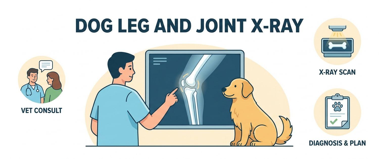
A dog starts limping and the first thing the clinic does is take a film. A leg and joint x-ray is cheaper, faster and more available than anything else, and it answers half the questions on the spot. Here is what it shows, why positioning decides everything in dysplasia, and where the limits of plain film lie.
What a Dog Leg and Joint X-Ray Shows
An x-ray sees dense things: bone, joint spaces, mineralisation, metal. So it answers questions such as:
- is there a fracture, a crack, a luxation or a subluxation;
- how the joint space looks — narrowed, widened, congruent or not;
- are there bony outgrowths (osteophytes), the mark of a long-standing process;
- is any bone being destroyed — always a reason for further work-up;
- are pins and plates sitting correctly after surgery.
Ligaments, menisci, cartilage and tendons are invisible on film. A cranial cruciate ligament tear — the commonest cause of hind-limb lameness — shows only indirectly, through joint effusion and displacement. The general logic of plain films is covered in dog X-ray.
Canine Hip Dysplasia on X-Ray
Dysplasia is a mismatch between the femoral head and the socket. The film is read for how deeply the head sits, whether it is subluxated and whether the joint contour has changed.
The part owners rarely hear: dysplasia can only be graded on a strictly symmetrical film. The dog lies on its back, hind limbs extended and rotated inward, parallel to each other. The smallest pelvic tilt changes the picture — a healthy joint can look subluxated and a bad one almost normal. That is why official dysplasia films are taken under sedation: a conscious animal cannot be held in that position.
Screening films are usually taken after the growth plates close; the exact age depends on breed and grading scheme, and the vet sets it. Elbow dysplasia is graded differently and needs its own views.
Fractures and Dislocations: What Is Visible at Once
A fresh break shows as a lucent line breaking the bone contour. The report describes its location, pattern (transverse, oblique, comminuted), displacement and whether the joint is involved. Hairline fractures sometimes do not show on day one and appear 7–10 days later as the bone remodels, so a repeat film is taken if pain persists.
Puppies have their own trap: growth-plate fractures look deceptively minor and have serious consequences for limb length.
Dog Spine X-Ray: Discs and Vertebrae
A spinal film shows the shape and number of vertebrae, the width of the disc spaces, spondylosis, fractures and congenital anomalies such as wedge vertebrae in flat-faced breeds. What it does not show is the spinal cord itself or disc material: a narrowed space only hints at a herniation and says nothing about how badly the cord is compressed. Only cross-sectional imaging answers that — see MRI and CT in dogs and cats.
Arthritis and Age-Related Changes
In an older dog a film almost always shows something: osteophytes, subchondral sclerosis, a narrowed space. What matters is that the amount of arthritis on the film matches pain poorly. A dog with obvious changes may run happily, while one with minimal changes limps badly. So the film is read together with the exam and the gait, a fork described in dog limping.
How a Dog X-Ray Is Done and Why Sedation Helps
The procedure takes minutes: the dog is placed on the table and at least two views are taken, front and side, because a single projection cannot show volume.
Sedation is not always needed, but it is offered when:
- strict positioning is required (hips, elbows, spine);
- the dog is in pain and resists any movement of the limb;
- the animal is large and cannot be held safely.
Sedation is not full anaesthesia, but it is not trivial either: the vet examines the dog first and often asks for blood work. What those numbers mean is covered by pet results interpretation and the dog blood test article. Holding the animal by hand during exposure is discouraged: the person takes the dose and the positioning still comes out worse.
Reading the X-Ray Report
| Phrase | What it usually means |
|---|---|
| Joint surfaces congruent | The joint fits together — normal |
| Subluxation of the femoral head | The head is partly out of the socket — the dysplasia picture |
| Osteophytes at the joint margins | Bony outgrowths, a sign of arthritis |
| Narrowed joint space | Thinning cartilage, most often age-related |
| Fracture line without displacement | The bone is broken but the fragments are in place |
| Zone of bone lysis | Bone destruction — needs urgent clarification |
| Lumbosacral spondylosis | Bony bridges between vertebrae, a common age finding |
Clinics may hand out a disc or DICOM files. If they will not open, see how to open an MRI or CT disc and how to open a DICOM (.dcm) file.
When Plain Films Are Not Enough
If the film is clean and the limp persists, the search moves to soft tissue and nerves: joint ultrasound, CT for complex bone anatomy, MRI for a suspected disc or nerve. In large animals the same logic applies with a size correction, as described in horse X-ray and ultrasound.
If you already hold the film, upload it or a photo of the report to pet imaging interpretation: the service explains the wording in plain language and builds the questions to ask. If there is no film yet, only a changed gait, it is worth using the dog symptom checker or simply describing the symptoms.
This article is informational. Diagnosis and treatment of your pet are the job of a veterinarian.
For informational purposes only
This article is for informational purposes only and does not constitute medical advice, diagnosis, or treatment. Please consult a healthcare professional for medical guidance.