Fluorography: What It Shows and How It Differs from X-ray
Reviewed by the LabReadAI medical team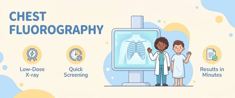
Fluorography is a mass chest screening that in Russia is done once a year. The report says "lung fields without focal shadows", "roots are structured", "sinuses are clear" — and the person cannot tell whether all is well. Let's break down what fluorography shows, how it differs from X-ray and what the common phrases mean.
What Fluorography Shows and Why It Is Done
Fluorography is a screening chest X-ray image. Its main purpose is mass screening: to catch signs of tuberculosis, large focal lung lesions and gross heart and pleural disease in time. The method shows the overall picture of the lung fields, the lung roots, the heart contour and the diaphragm — but it is screening, not detailed diagnosis.
How Fluorography Differs from X-ray and CT
All three methods use X-rays but differ in detail:
- Fluorography — fast, cheap screening at a low dose; lower resolution, small details can be missed.
- Radiography (X-ray) — a higher-quality image; ordered specifically when fluorography raises suspicion or symptoms are present.
- CT — the most detailed method, sees small lesions; higher dose, used for clarification.
So fluorography answers "is there gross pathology", while X-ray and CT answer "what exactly and how serious". Other regions use fundamentally different methods — for example MRI for soft tissue or ultrasound for abdominal organs.
How Often It Is Done and Who Should Not
For prevention, fluorography is usually done once a year; the dose with digital fluorography is small. It is not performed in pregnancy (except emergencies), and mass screening in children is replaced by other methods. With symptoms (a cough lasting over 3 weeks, coughing up blood, weight loss), do not wait for the scheduled date — see a doctor, who will order a targeted image.
Common Conclusions: Roots, Sinuses, Lung Markings
| Phrasing | What it usually means |
|---|---|
| Lung fields without focal or infiltrative shadows | No gross pathology detected (normal) |
| Roots structured, not enlarged | Normal |
| Sinuses clear | No fluid in the pleural cavities (normal) |
| Increased lung markings | Non-specific: bronchitis, past infection, smoking |
| Roots enlarged/dense | Needs clarification (X-ray/CT) |
| Fibrotic changes, adhesions | Most often traces of old processes |
"Increased lung markings" and "fibrotic changes" are the most common "scary" phrases, which usually reflect residual or age-related changes rather than active disease.
What Fluorography Detects and What It Misses
Fluorography reliably notices large changes: signs of tuberculosis, big lesions, marked pleural effusion, a substantially enlarged heart. But because of its modest resolution it can miss small nodules and early stages — so with symptoms or risk factors, a normal fluorography does not replace X-ray or CT. It is a screening tool, not a way to rule disease out.
A "Suspicious" Fluorography: What Next
If the report shows a focal shadow, infiltration, enlarged roots or a mass, the doctor will order further imaging: a targeted two-view X-ray or a chest CT, sometimes blood tests and a pulmonologist's or TB specialist's review. The phrase "requires further evaluation" is not a diagnosis but a prompt to clarify.
How to Tell That Fluorography Is Normal
Signs of a normal report: lung fields without focal or infiltrative shadows, structured and non-enlarged roots, clear sinuses, a non-enlarged heart shadow and a normally positioned diaphragm. If all of these are present, no gross pathology was detected on screening.
You can upload a fluorography report or image and get a plain-language breakdown with the imaging interpretation service — it helps you understand the phrasing and separate residual changes from those that need further evaluation. The service does not replace a doctor.
This article is for informational purposes. Reading an image and the diagnosis are the doctor's job.
For informational purposes only
This article is for informational purposes only and does not constitute medical advice, diagnosis, or treatment. Please consult a healthcare professional for medical guidance.