Abdominal Ultrasound: What It Shows, Preparation and Results
Reviewed by the LabReadAI medical team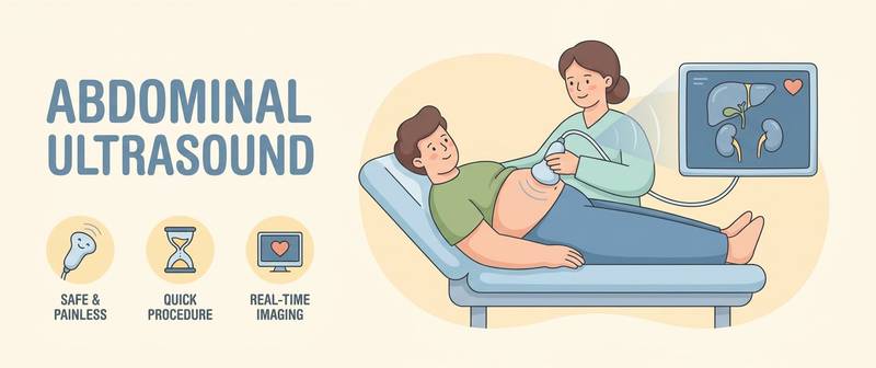
An abdominal ultrasound is one of the most common and safest investigations: no radiation, no pain, and in 15–20 minutes the doctor examines several organs at once. But the report hands the patient sizes in millimetres and words like "echogenicity", "diffuse changes", "homogeneous structure" — with no sense of whether that is normal. Let's break down what an abdominal ultrasound shows, how to prepare for it and how to read the result.
What an Abdominal Ultrasound Shows and Which Organs Are Assessed
Ultrasound shows the structure of organs: their size, shape, tissue density (echogenicity), contours, masses, fluid and stones. A standard scan covers:
- liver — size, echogenicity, focal lesions;
- gallbladder and ducts — walls, stones, bile stasis;
- pancreas — contours, structure, duct;
- spleen — size;
- kidneys — often examined together with the abdomen.
Know the limit of the method: ultrasound sees structure well but does not assess organ function. Fatty liver appears as a "bright" liver, but how impaired its work is can only be shown by blood tests (ALT, AST, bilirubin). So ultrasound and biochemistry complement each other.
Preparation for an Abdominal Ultrasound: Diet and What to Avoid
What the doctor can see depends directly on preparation. Bowel gas "shadows" the pancreas and vessels, and food contracts the gallbladder — so stones in it can be missed.
- Fasting — the last meal 8–12 hours before the scan (for adults). That is why abdominal ultrasound is usually done in the morning.
- For 2–3 days — a diet without gas-forming foods: avoid legumes, dark bread, cabbage, fizzy drinks, milk, and large amounts of raw vegetables and fruit.
- If prone to bloating, the doctor may suggest anti-flatulence agents or sorbents.
- You may drink water, but avoid drinking a lot right before the scan (unless a full bladder is needed to view the pelvis).
Good preparation is half the accuracy of the report. In an emergency (acute abdominal pain) the scan is done without preparation, and some detail may be unavailable.
Liver and Spleen: Norms and Common Findings
A healthy liver has smooth contours, a homogeneous structure and echogenicity similar to the renal cortex. Common report phrases:
- "Increased echogenicity / diffuse changes consistent with fatty liver" — fat accumulation in liver cells. This is the most common finding; more in fatty liver disease.
- "Nodular contour, heterogeneous structure, signs of portal hypertension" — may point to liver cirrhosis and warrants work-up.
- Focal lesions (cyst, haemangioma, nodule) — size and character are described; some are harmless, some need clarification on MRI/CT.
The spleen is assessed by size: enlargement (splenomegaly) can occur with blood disorders, infections and portal hypertension.
Gallbladder and Ducts: Stones and Stasis
The gallbladder is one of the main "stars" of ultrasound, because it shows stones extremely well. The report may mention:
- calculi (stones) — the key sign of gallstone disease; their number and size are described;
- wall thickening — a sign of inflammation (cholecystitis);
- bile stasis, a kinked gallbladder — common functional features.
It is for the gallbladder's sake that arriving strictly fasting matters: after eating it contracts and small stones are easily missed.
The Pancreas on Ultrasound
The pancreas is the hardest to image — bowel gas often covers it, so preparation is critical. Size, contour clarity and structure are assessed. Swelling and blurred contours can occur with inflammation — acute pancreatitis; the diagnosis is confirmed with blood enzymes (amylase, lipase). Diffuse changes and increased density more often reflect a chronic process or age-related change.
Kidneys and What Else Is Covered
The kidneys are often scanned with the abdomen. Ultrasound shows size, structure, dilation of the collecting system and stones — when kidney stones are suspected. Free fluid in the abdomen and large vessels (the aorta) are also assessed.
Common Ultrasound Conclusions and What They Mean
| Phrasing | What it usually means |
|---|---|
| Diffuse changes of liver/pancreas | Non-specific sign — assessed with blood tests |
| Increased liver echogenicity | Most often fatty liver |
| Gallbladder calculi | Stones — gallstone disease |
| Kinked gallbladder | Usually a functional feature, not a disease |
| Microliths / sand in kidneys | Tiny inclusions, monitored |
| Hepatomegaly / splenomegaly | Enlarged liver/spleen — the cause must be sought |
"Diffuse changes" is the most alarming yet most non-specific phrase: it is not a diagnosis but a description, always interpreted together with blood tests and symptoms.
When Tests and a Doctor Are Needed: Limits of the Method
Ultrasound shows a picture but does not make a final diagnosis or assess function. After it, the following are often needed:
- blood biochemistry (liver panel, pancreatic enzymes);
- for focal lesions — MRI or CT for clarification;
- a gastroenterologist or surgeon for stones and inflammation.
You can upload your ultrasound report or image and get a plain-language breakdown with the imaging and ultrasound interpretation service — it helps you understand what is normal in the protocol and what to discuss with a doctor. The service does not replace an in-person exam.
This article is for informational purposes. The final reading of an ultrasound and the diagnosis are the doctor's job.
For informational purposes only
This article is for informational purposes only and does not constitute medical advice, diagnosis, or treatment. Please consult a healthcare professional for medical guidance.