AST in Blood: Normal Levels, Causes of Elevation and Interpretation
Reviewed by the LabReadAI medical team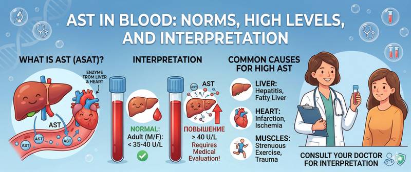
AST and ALT are almost always measured together. But there is a fundamental difference between them that changes the diagnostic logic: while ALT is predominantly a liver enzyme, aspartate aminotransferase (AST) is distributed much more widely. It is present at high concentrations in heart muscle cells, skeletal muscle and the liver simultaneously. This makes AST less specific for the liver, but more informative when the question is not whether damage has occurred — but where it is coming from. Here is how to read this marker correctly.
What Aspartate Aminotransferase (AST) Is and How It Differs from ALT
AST (aspartate aminotransferase) belongs to the transaminase enzyme family. It transfers an amino group from aspartate to alpha-ketoglutarate — a reaction that links amino acid metabolism to the Krebs cycle. The enzyme exists in two cellular pools: cytoplasmic (the smaller fraction) and mitochondrial (the larger fraction, around 80% of the total).
This dual localisation explains a clinically important feature of AST. Mild cell injury disrupts only the outer membrane — cytoplasmic AST is released. Severe injury, particularly from alcohol, destroys mitochondria as well — mitochondrial AST pours out in large quantities. This is why AST is disproportionately elevated compared to ALT in alcoholic hepatitis.
The key distinction from ALT is tissue specificity. AST is present at high concentrations in:
- Cardiomyocytes — heart muscle cells
- Hepatocytes — liver cells
- Skeletal muscle cells
- To a lesser extent — kidneys, pancreas, and erythrocytes
This is why isolated AST elevation with a normal ALT should direct attention to the heart or muscles rather than the liver. Conversely, parallel elevation of both enzymes almost invariably points to hepatocellular injury.
Normal AST Levels in Blood
AST reference values, like those for ALT, depend on sex and age.
| Group | AST normal range, U/L |
|---|---|
| Men 18–60 years | up to 40 |
| Men over 60 years | up to 35 |
| Women 18–60 years | up to 32 |
| Women over 60 years | up to 30 |
| Children under 1 year | up to 58 |
| Children 1–14 years | up to 46 |
Reference ranges at your specific laboratory may differ slightly. Always use the values printed on your own lab report.
In children, AST norms are somewhat higher than in adults — physiologically driven by active growth and tissue remodelling. In newborns during the first days of life, AST can reach 100–140 U/L — a normal finding related to perinatal stress.
How to Prepare for an AST Blood Test
AST is measured from venous blood as part of a biochemistry panel.
Fasting. Strictly fasting — last meal 8–12 hours before. Fatty food has a minor direct effect on AST but standardising conditions is important for comparing results over time.
Exercise. Particularly critical for AST: skeletal muscles contain it at high concentrations. Intense strength training, long-distance running or even intramuscular injections can raise AST 2–5 fold. Avoid strenuous activity for 48 hours before the test.
Alcohol. Avoid for 48–72 hours — alcohol directly damages hepatocyte mitochondria, the very AST pool released in alcoholic hepatitis.
Haemolysis. Erythrocytes contain AST. If haemolysis occurs in the sample tube during incorrect storage or transport, the result will be falsely elevated. Quality laboratories flag this in the report.
Medications. Statins, antibiotics, NSAIDs, isoniazid, amiodarone and several other drugs raise AST. Inform your doctor of any medications you are taking.
Elevated AST: Causes and Degrees of Elevation
The key diagnostic question when AST is elevated is: where is it coming from — liver, heart or muscle? The answer usually lies in the pattern of accompanying markers rather than the absolute value alone.
| Degree of elevation | Fold above ULN | Typical causes |
|---|---|---|
| Mild | 1–3× ULN | NAFLD, moderate alcohol, drug effect, intense exercise, hypothyroidism |
| Moderate | 3–10× ULN | Chronic hepatitis B/C, alcoholic hepatitis, autoimmune hepatitis, myositis, mild rhabdomyolysis |
| High | 10–25× ULN | Acute viral hepatitis, severe alcoholic hepatitis, myocardial infarction, marked rhabdomyolysis |
| Very high | > 25× ULN | Fulminant hepatitis, massive myocardial infarction, massive rhabdomyolysis, acute hepatic ischaemia |
The absolute AST value does not equal the severity of damage. As with ALT, in end-stage cirrhosis AST may be only mildly elevated because so few viable cells remain to release it.
Three main sources of AST elevation and how to distinguish them:
Hepatic source — AST rises in parallel with ALT; the de Ritis ratio is variable; GGT and bilirubin are often elevated. Liver function panel and ultrasound confirm the source.
Cardiac source — AST is elevated with normal or only mildly raised ALT; GGT is normal. Historically, AST was the first cardiac marker used in myocardial infarction — it begins rising 6–8 hours after ischaemia, peaks at 24–36 hours, and normalises within 4–5 days. It has been entirely superseded by troponin, which is far more specific and appears much earlier. In suspected infarction, AST has no independent diagnostic role.
Muscular source — AST elevated with normal ALT and GGT. Typically linked to physical activity, trauma, seizures or statin use. Confirmed by simultaneous elevation of CK (creatine kinase), which is far more specific for muscle damage.
Low AST
AST below the reference range is uncommon and of limited clinical significance. It occurs in:
- Vitamin B6 deficiency — pyridoxal phosphate is a cofactor for transaminase activity; its deficiency reduces enzyme activity
- End-stage hepatic insufficiency — by the same logic as low ALT: no cells left to produce the enzyme
- Metronidazole and certain other drugs — through competition at the enzyme's active site
AST and ALT: The De Ritis Ratio in Clinical Practice
The AST:ALT ratio is one of the most practical diagnostic tools in hepatology. It is calculated simply by dividing the AST value by the ALT value.
| AST:ALT ratio | Diagnostic significance |
|---|---|
| < 0.8 | Viral hepatitis, NAFLD, drug-induced liver injury — ALT dominates |
| 0.8–1.2 | Non-specific; requires additional contextual analysis |
| > 2.0 | Alcoholic hepatitis — high specificity when GGT is also elevated |
| > 3.0 | Virtually pathognomonic for alcoholic hepatitis or rhabdomyolysis |
An important caveat: the de Ritis ratio is only informative when both enzymes are simultaneously elevated. Calculating the ratio when both values are within normal range is meaningless. The ratio also loses diagnostic value at very high absolute levels (> 1000 U/L), where any cause of massive cytolysis can produce a similar picture.
For a complete assessment, both enzymes are reviewed as part of a liver function test panel together with GGT, alkaline phosphatase, bilirubin and albumin — only this combination allows hepatocellular to be distinguished from cholestatic injury.
High AST — What Does It Mean and When to See a Doctor Urgently
- AST above 500 U/L — regardless of symptoms; requires urgent assessment and source identification
- AST elevated with chest pain, breathlessness or palpitations — seek emergency care immediately: possible acute coronary syndrome, though diagnosis is confirmed with troponin
- Rising AST on serial testing — even at moderate levels, a consistent upward trend warrants investigation
- High AST with dark urine (tea- or cola-coloured) — sign of myoglobinuria in rhabdomyolysis; risk of acute kidney injury
- Jaundice with high AST — significant hepatic injury with functional impairment
On an elective basis: any AST elevation above normal found incidentally warrants a GP visit without delay. AST cannot be interpreted in isolation — it only tells its full story alongside ALT, GGT and the rest of the liver function panel.
This article is for informational purposes only. Interpretation of test results and diagnosis are the responsibility of a qualified physician.
For informational purposes only
This article is for informational purposes only and does not constitute medical advice, diagnosis, or treatment. Please consult a healthcare professional for medical guidance.