Kidney Stones: Symptoms, Causes, Treatment and Prevention
Reviewed by the LabReadAI medical team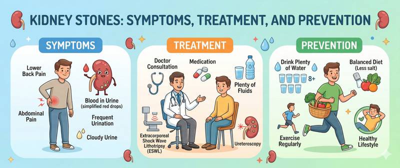
The pain of renal colic is described as among the most intense in clinical medicine — sharp, cramping, radiating from the flank to the groin, offering no position of relief. This is how a stone that has left the kidney travels through the ureter. Kidney stones affect 10–15% of adults worldwide, recur in every second patient within 10 years, and remain one of the leading causes of emergency urology admissions.
What Are Kidney Stones and How Do They Form
Kidney stones (nephrolithiasis, urolithiasis) are hard crystalline deposits forming in the kidneys, ureters, bladder, or urethra as a result of changes in the chemical composition of urine. When the concentration of certain substances exceeds their solubility threshold, they begin to crystallise and gradually aggregate into stones.
Stone formation takes months or years. A microscopic crystal acts as a "nucleus" onto which new layers deposit. While a stone remains in the renal pelvis without obstructing flow, it may cause no symptoms at all — sometimes for years. Pain begins the moment the stone moves and lodges in the ureter, blocking urine drainage.
Three core conditions are required for stone formation:
- urine supersaturation — excessively high concentrations of stone-forming substances (oxalate, calcium, uric acid, phosphate);
- insufficient urine volume — low fluid intake raises concentrations;
- deficiency of inhibitors — citrate, magnesium, and other substances normally prevent crystallisation; their reduction creates conditions for stone growth.
Causes and Risk Factors for Kidney Stones
Insufficient fluid intake — the most common and most correctable factor. When daily urine output falls below 1 litre, stone-forming risk is maximal. Hot climates and intense physical activity without fluid replacement produce the same effect.
Diet. Excess animal protein increases urinary excretion of uric acid, oxalate, and calcium. High salt intake raises urinary calcium excretion. Large amounts of oxalate-rich foods (spinach, beets, nuts, chocolate) raise the risk of calcium oxalate stones.
Metabolic abnormalities:
- Elevated uric acid — the primary risk factor for urate stones; frequently accompanies hyperuricaemia and gout.
- Elevated blood calcium — in hypercalcaemia and hyperparathyroidism, excess calcium is filtered into the urine and precipitates stones.
- Hyperoxaluria — primary (genetic) or secondary (malabsorption, celiac disease, bowel resection).
- Urinary citrate deficiency — citrate binds calcium and prevents crystallisation; its reduction is typical in chronic acidosis and persistent diarrhoea.
Systemic conditions:
- Chronic kidney disease disrupts regulation of calcium and phosphate excretion.
- Arterial hypertension — an independent risk factor, partly through impaired renal calcium reabsorption.
- Obesity and type 2 diabetes — lower urinary pH (acidic urine favours urate stones) and increase oxalate excretion.
- Crohn's disease and ileal resection — impaired fat absorption causes intraluminal calcium binding, increasing oxalate absorption.
Anatomical factors: structural abnormalities of the collecting system (horseshoe kidney, ureteral strictures) promote urine stasis and stone formation.
Heredity. A family history of kidney stones raises individual risk two- to three-fold. Rare genetic conditions — cystinuria, primary hyperoxaluria — produce recurrent stones from childhood.
Symptoms of Kidney Stones: Renal Colic and Other Presentations
The clinical picture depends on stone size, location, and whether obstruction is present.
Stone in the renal pelvis without obstruction — usually asymptomatic. Large staghorn calculi are often found incidentally on ultrasound. The only complaint may be a dull persistent flank ache.
Renal colic — acute ureteral obstruction. The classic picture:
- severe, excruciating cramping flank pain radiating along the ureter toward the groin, inner thigh, and genitals;
- the pain is colicky — it waxes and wanes but never fully resolves — and the patient cannot find a comfortable position (in contrast to peritoneal pain, where movement worsens everything);
- nausea and vomiting — reflex in origin;
- frequent, painful urination when the stone is in the lower third of the ureter;
- blood in the urine (haematuria) — visible or microscopic.
Stone size predicts outcome: stones up to 4–5 mm pass spontaneously in 80–90% of cases; 5–7 mm in approximately 50%; above 7–8 mm they almost always require instrumentation or surgery.
Infected stone — the most dangerous scenario. Ureteral obstruction combined with urinary tract infection creates the risk of obstructive pyelonephritis and urosepsis: high fever, rigors, escalating flank pain. This is an indication for emergency renal drainage.
Chronic course — recurrent colic episodes, chronic dull flank pain, recurrent urinary tract infections, gradual decline in renal function.
Types of Kidney Stones by Chemical Composition
Stone composition determines its cause, treatment, and prevention strategy.
| Stone type | Prevalence | CT density | Key cause |
|---|---|---|---|
| Calcium oxalate | 60–70% | Dense, white | Hyperoxaluria, hypercalciuria |
| Calcium phosphate | 10–20% | Very dense | Hyperparathyroidism, renal tubular acidosis |
| Urate | 10–15% | Radiolucent (invisible on X-ray) | High uric acid, acidic urine |
| Struvite | 5–10% | Staghorn shape | Chronic urease-producing infection |
| Cystine | < 1% | Mildly dense | Cystinuria (genetic disorder) |
Urate stones are the only type that can be dissolved conservatively — by alkalinising urine to pH 6.5–7.0 with citrate preparations — without any surgical procedure.
Struvite (staghorn) stones form in chronic infection by urease-producing bacteria (Proteus, Klebsiella); they fill the entire renal pelvis and almost never pass spontaneously.
Diagnosing Kidney Stones
Imaging:
- Non-contrast CT of the abdomen (NCCT) — the gold standard in acute renal colic. Detects stones of any composition (except rare drug-induced calculi), determines size, location, and degree of obstruction. Sensitivity 97–98%.
- Renal ultrasound — first-line in surveillance and in pregnancy. Identifies stones in the renal pelvis and collecting system dilatation as an indirect obstruction sign; poor sensitivity for ureteral stones.
- Plain abdominal X-ray — visualises only radiopaque stones (oxalate, phosphate); urate and cystine stones are invisible.
Laboratory workup — essential minimum:
- Urinalysis — red cells (haematuria), white cells (infection), crystals, urine pH. Acidic pH (< 5.5) is typical for urate stones; alkaline (> 7.0) for struvite.
- Kidney function test — creatinine and eGFR assess the degree of obstruction-related renal impairment.
- Serum uric acid, calcium, and phosphate — identify metabolic drivers.
Extended metabolic workup (for recurrent stones and young patients):
- 24-hour urine for calcium, oxalate, citrate, uric acid, and volume — identifies the specific metabolic defect.
- Parathyroid hormone — excludes primary hyperparathyroidism.
- Stone composition analysis — more informative than any blood test in establishing the cause.
Treatment of Kidney Stones
Conservative management (stones up to 5–6 mm):
- Liberal fluid intake — accelerates stone passage; target daily urine output ≥ 2–2.5 litres.
- Analgesia — NSAIDs (diclofenac, ketorolac) are first-line: they relieve ureteral spasm and mucosal oedema. Opioids when NSAIDs are insufficient.
- Alpha-blockers (tamsulosin) — relax the smooth muscle of the lower ureter, facilitating spontaneous passage.
- Urine alkalinisation (potassium citrate, sodium bicarbonate) — effective for urate stones.
Instrumentation and surgery:
- Extracorporeal shock wave lithotripsy (ESWL) — non-invasive fragmentation by external shock waves; suitable for stones up to 15–20 mm in the renal pelvis and upper ureter.
- Ureteroscopy with laser lithotripsy (URS) — an endoscope is passed through the urethra to the stone; a laser fragments it. Effective for ureteral stones at any level.
- Percutaneous nephrolithotomy (PCNL) — percutaneous renal access to remove large (> 20 mm) or staghorn calculi.
- Laparoscopic and open surgery — reserved for complex anatomical cases or failed minimally invasive approaches.
Pharmacological relapse prevention is tailored to metabolic workup findings:
- hyperuricosuria — allopurinol reduces uric acid production;
- hyperoxaluria — pyridoxine and dietary oxalate restriction;
- hypercalciuria — thiazide diuretics reduce urinary calcium excretion;
- low citrate — potassium citrate supplementation.
Prevention and When to See a Doctor
Universal prevention measures that work for any stone type:
- Fluid: drink at least 2.5–3 litres per day, aiming for urine output ≥ 2–2.5 litres. Water is the foundation; moderate amounts of lemon juice add urinary citrate. Sugar-sweetened drinks and grapefruit juice increase risk.
- Limit salt to 4–5 g/day — reduces urinary calcium excretion.
- Moderate animal protein — no more than 0.8–1.0 g/kg/day.
- Maintain normal dietary calcium intake (1000–1200 mg/day) — do not restrict, as intestinal calcium binds oxalate and reduces its absorption. Restricting calcium paradoxically raises the risk of oxalate stones.
- Maintain a healthy weight — obesity independently increases recurrence risk regardless of stone type.
See a urologist if:
- you have had a first episode of renal colic — even if the stone passed spontaneously;
- stones have recurred — full metabolic workup is indicated;
- stones occur in children or young adults (under 30) — a genetic or metabolic cause is highly likely;
- bilateral stones or a stone in a solitary kidney;
- progressive decline in kidney function on blood tests.
Call emergency services immediately if flank pain is accompanied by high fever and rigors — these are signs of obstructive pyelonephritis, a life-threatening condition requiring emergency renal drainage. Do not interpret your test results on your own; consult a urologist for treatment and relapse prevention planning.
For informational purposes only
This article is for informational purposes only and does not constitute medical advice, diagnosis, or treatment. Please consult a healthcare professional for medical guidance.