High Blood Calcium (Hypercalcaemia): Causes and Treatment
Reviewed by the LabReadAI medical team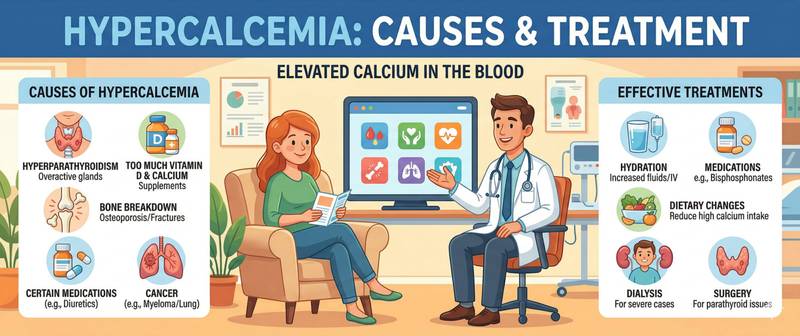
Finding "calcium elevated" on a blood test report is a signal that should not be put off until the next appointment. Calcium in blood governs nerve conduction, muscle contraction, blood clotting and bone mineral density. When levels exceed the normal range, the body sends a broad spectrum of signals — from persistent fatigue and thirst to dangerous cardiac arrhythmias. This article explains what hypercalcaemia is, why it develops and what steps to take when blood calcium is high.
What Is Hypercalcaemia and What Are Normal Blood Calcium Levels
Hypercalcaemia is defined as a serum total calcium above 2.6 mmol/L, or an ionised calcium above 1.3 mmol/L. The normal range for adults is: total calcium 2.15–2.55 mmol/L, ionised calcium 1.15–1.30 mmol/L.
Severity is classified in three tiers:
| Grade | Total calcium | Clinical significance |
|---|---|---|
| Mild | 2.6–3.0 mmol/L | Often asymptomatic, found incidentally |
| Moderate | 3.0–3.5 mmol/L | Symptoms present, treatment required |
| Severe | > 3.5 mmol/L | Life-threatening, emergency care needed |
Mild hypercalcaemia is frequently discovered on a routine biochemistry panel when the patient feels perfectly well. Moderate and severe forms produce a clear clinical picture that must not be missed.
Symptoms of High Blood Calcium
The classic symptom complex is captured by the medical mnemonic bones, stones, groans and psychic moans, reflecting four main systemic effects of excess calcium.
Bones: bone and joint pain, pathological fractures from minor trauma as calcium leaches from the skeleton into the bloodstream.
Stones: the kidneys try to excrete the calcium overload, leading to oxalate and phosphate kidney stones — flank pain, haematuria and recurrent renal colic.
Groans (gastrointestinal): nausea, vomiting, constipation, loss of appetite and abdominal pain. Excess calcium inhibits smooth muscle throughout the gut.
Psychic moans (neuropsychiatric): fatigue, lethargy, difficulty concentrating, low mood, confusion. In severe hypercalcaemia, coma is possible.
Cardiovascular: shortening of the QT interval on ECG, bradycardia, risk of arrhythmias. In patients taking cardiac glycosides, hypercalcaemia greatly amplifies toxicity.
Polyuria and polydipsia: frequent urination and persistent thirst — excess calcium blunts the kidney's response to antidiuretic hormone.
Causes of Hypercalcaemia: From Hyperparathyroidism to Cancer
Over 90% of hypercalcaemia cases are explained by just two causes — primary hyperparathyroidism and malignancy.
Primary hyperparathyroidism is the most common cause in outpatient settings. One or more parathyroid glands begin autonomously producing excess parathyroid hormone, instructing bones to release calcium and the kidneys to retain it. A benign adenoma is the usual culprit. For a detailed look at the condition, see primary hyperparathyroidism.
Malignancy is the leading cause among hospitalised patients. Tumours may secrete PTH-related protein (PTHrP), directly destroy bone, or synthesise active vitamin D. Hypercalcaemia is especially common in multiple myeloma, as well as lung, breast and renal cancers.
Excess vitamin D. Uncontrolled high-dose vitamin D supplementation enhances intestinal calcium absorption — one reason why self-prescribing megadoses is risky. For more on vitamin D imbalance, see vitamin D deficiency.
Sarcoidosis and granulomatous diseases. Activated macrophages within granulomas synthesise calcitriol (active vitamin D), increasing calcium absorption from the diet.
Other causes: prolonged immobilisation (calcium mobilises from inactive bone), thiazide diuretics, familial hypocalciuric hypercalcaemia — a rare inherited condition with a benign course.
How Hypercalcaemia Is Diagnosed: Which Tests Are Needed
Detecting high calcium is only the first step. The key task is finding the cause. The standard diagnostic algorithm proceeds as follows.
Step 1 — confirm hypercalcaemia. A single elevated result may be artefactual: a prolonged tourniquet, haemolysis or a high albumin level all inflate total calcium. Total and ionised calcium are repeated, and albumin is measured for corrected calcium calculation.
Step 2 — parathyroid hormone (PTH). This is the pivotal differential test. High PTH with high calcium → primary hyperparathyroidism. Low PTH with high calcium → malignancy, vitamin D toxicity, sarcoidosis.
Step 3 — additional markers:
| Test | Purpose |
|---|---|
| Blood phosphorus | Low in hyperparathyroidism; elevated in renal failure |
| Creatinine | Renal function assessment — calcium is nephrotoxic |
| Vitamin D (25-OH) | Rule out hypervitaminosis D |
| PTH-related protein (PTHrP) | Marker of paraneoplastic hypercalcaemia |
A full kidney function test is mandatory in any hypercalcaemia — the kidneys are the first organ damaged by sustained calcium overload.
Complications of Hypercalcaemia
Chronic hypercalcaemia, even mild, causes systemic damage over time.
Kidneys. Nephrocalcinosis — calcium deposits in renal tissue — leads to progressive kidney failure. The acute form is acute kidney injury triggered by severe hypercalcaemia combined with dehydration.
Bones. Prolonged stimulation of bone resorption causes osteoporosis and pathological fractures. The paradox: blood calcium is high while the skeleton is being depleted.
Heart. A shortened QT interval raises the risk of ventricular arrhythmias. Patients on digoxin are particularly vulnerable, as hypercalcaemia multiplies glycoside toxicity.
Gastrointestinal tract. Elevated calcium stimulates gastrin secretion, raising the risk of peptic ulcers. Chronic constipation significantly impairs quality of life.
Treatment of Hypercalcaemia
Treatment depends on severity and the underlying cause.
Mild hypercalcaemia (2.6–3.0 mmol/L) due to asymptomatic primary hyperparathyroidism in older patients may need only surveillance: calcium monitoring every 6–12 months, adequate fluid intake (at least 2 litres daily) and moderate weight-bearing exercise, which slows bone resorption.
Moderate and severe hypercalcaemia requires active intervention:
- Aggressive IV hydration with isotonic saline — the first and most critical step. Restoring circulating volume enhances renal calcium excretion.
- Bisphosphonates (zoledronic acid, pamidronate) — suppress bone resorption. Effect develops within 48–72 hours.
- Calcitonin — rapidly lowers calcium but the effect is short-lived.
- Glucocorticoids — effective for hypercalcaemia caused by sarcoidosis, other granulomatoses and haematological malignancies.
- Dialysis — in severe renal failure when other measures are insufficient.
In symptomatic primary hyperparathyroidism, surgical removal of the adenoma offers definitive, long-lasting cure.
When High Calcium Is a Medical Emergency
Hypercalcaemic crisis is a life-threatening condition that develops when calcium exceeds 3.5 mmol/L, often triggered by dehydration or acute illness. Signs requiring immediate emergency care:
- sudden severe weakness, confusion or disorientation
- intractable vomiting with progressive dehydration
- cardiac arrhythmias or haemodynamic instability
- oliguria — markedly reduced urine output
Self-treatment and watchful waiting are not options in this situation. Even a moderate asymptomatic rise in calcium is reason to see an endocrinologist for full investigation — not simply to "repeat the test next month."
Key Takeaways
Hypercalcaemia is always a symptom, not a disease in itself. Behind elevated calcium may be a benign parathyroid adenoma or an early-stage malignancy. That is why any result above the normal range warrants thorough investigation — with PTH, phosphorus and a renal function assessment. The earlier the cause is found, the more effective the treatment and the better the long-term outcome.
This article is for informational purposes only. Interpretation of test results and treatment decisions should be made by a qualified healthcare professional.
For informational purposes only
This article is for informational purposes only and does not constitute medical advice, diagnosis, or treatment. Please consult a healthcare professional for medical guidance.