Gallstone Disease: Symptoms, Causes, Diagnosis and Treatment
Reviewed by the LabReadAI medical team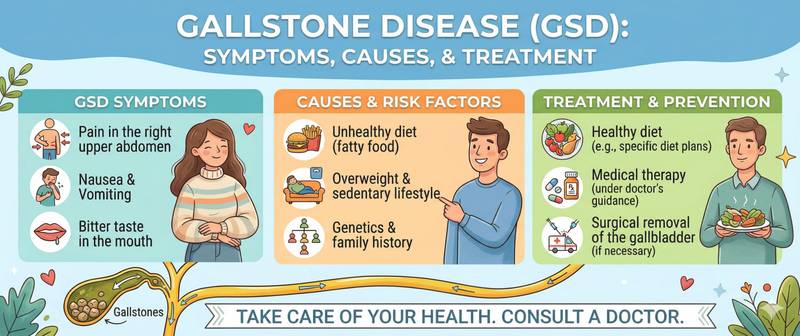
One in ten adults carries gallstones — and most have no idea. Gallstone disease can remain silent for decades, then without warning produce severe right upper quadrant pain in the middle of the night after a fatty dinner. Knowing how to recognise biliary colic, distinguish it from appendicitis, and understand when watchful waiting is appropriate versus when surgery is needed.
What Gallstone Disease Is and How Stones Form
Gallstone disease (cholelithiasis) is the formation of concretions (stones) in the gallbladder and bile ducts. Bile is a complex fluid: it holds cholesterol, bile acids, phospholipids, bilirubin, and mineral salts in solution. While all components remain in proper balance, bile stays liquid. When that balance is disrupted, crystallisation begins.
Types of stones:
Cholesterol stones (80–90% in Western countries) — form when bile is supersaturated with cholesterol. Yellowish-white, often solitary and large. Risk factors: obesity, rapid weight loss, pregnancy, female sex, oestrogen use, age > 40, sedentary lifestyle.
Pigment stones (10–20%) — formed from insoluble calcium-bilirubin salts. Black stones (in haemolysis, cirrhosis, Crohn's disease) and brown stones (in chronic biliary infection). In haemolytic anaemia, persistently elevated indirect bilirubin is a classic driver of pigment stone formation.
The "4 F" rule (Fat, Female, Forty, Fertile) — a classic mnemonic for risk factors, though gallstones occur in all groups.
Symptoms: From Silent Stones to Biliary Colic
Asymptomatic cholelithiasis — the most common presentation: 70–80% of people with gallstones never experience symptoms. Stones are found incidentally on ultrasound. The risk of developing symptoms is approximately 2% per year.
Biliary colic — acute episodic pain occurring when a stone temporarily obstructs the gallbladder outlet (neck or cystic duct).
Pain characteristics:
- Location: right upper quadrant or epigastrium
- Radiation: to the right shoulder, scapula, or interscapular area
- Character: intense, constant (despite the term "colic"), peaks within 15–30 minutes
- Provocation: fatty food, large meals, particularly in the evening
- Duration: 15 minutes to 6 hours. Pain lasting > 6 hours → suspect acute cholecystitis
- Associated: nausea, vomiting, occasionally low-grade fever
Dyspepsia — right upper quadrant heaviness after fatty food, bitter taste, bloating. Non-specific; often accompanies gallstone disease but can reflect other conditions.
Complications of Gallstone Disease
Untreated gallstone disease can produce life-threatening complications.
Acute cholecystitis — gallbladder inflammation, most commonly from stone obstruction. Pain lasts > 6 hours, fever, positive Murphy's sign (sharp pain on deep palpation under the right costal margin during inspiration), leucocytosis. Requires hospitalisation and often emergency cholecystectomy.
Choledocholithiasis — stone migration from the gallbladder into the common bile duct. Obstructive jaundice results: elevated direct bilirubin, ALP, GGT, acholic stools, dark urine, pruritus. Diagnosis: ultrasound (dilated common bile duct), MRCP, or ERCP.
Cholangitis — infection of an obstructed bile duct. Charcot's triad: fever with rigors + jaundice + right upper quadrant pain. Reynolds' pentad in severe cholangitis: + hypotension + altered consciousness. A life-threatening emergency requiring immediate biliary decompression (ERCP) and antibiotics.
Acute biliary pancreatitis — a stone impacted at the ampulla of Vater blocks the pancreatic duct. Severe epigastric pain radiating to the back; markedly elevated amylase and lipase. A leading cause of acute pancreatitis.
Gallbladder cancer — a rare but serious complication of longstanding gallstone disease. Large stones (> 3 cm) and porcelain gallbladder are risk factors.
Diagnosis of Gallstone Disease
Abdominal ultrasound — the gold standard: sensitivity > 95% for gallbladder stones. Identifies stones, wall thickness, duct dilation. Performed fasting (the gallbladder must be distended).
Laboratory findings:
In uncomplicated gallstone disease, blood tests may be entirely normal. With complications:
- Complete blood count: leucocytosis with neutrophilia — cholecystitis, cholangitis
- Direct bilirubin, ALP, GGT — elevated in choledocholithiasis and cholestasis
- ALT, AST — mildly elevated in acute cholestasis
- Amylase — markedly elevated in biliary pancreatitis
- C-reactive protein — inflammatory marker in cholecystitis and cholangitis
CT, MRCP — for suspected choledocholithiasis and pre-procedural planning.
ERCP (endoscopic retrograde cholangiopancreatography) — simultaneous diagnosis and treatment of choledocholithiasis: stones can be removed from the duct endoscopically.
Treatment of Gallstone Disease
Asymptomatic cholelithiasis: watchful waiting is generally appropriate. Prophylactic cholecystectomy is considered for: very large stones (> 3 cm), calcified ("porcelain") gallbladder, sickle cell anaemia (high crisis risk), patients undergoing bariatric surgery.
Symptomatic gallstone disease (biliary colic): cholecystectomy is the standard of care.
Laparoscopic cholecystectomy — the gold standard: minimally invasive (4 small incisions), discharge within 1–2 days, return to work within 1–2 weeks. Performed electively for recurrent colic.
Emergency cholecystectomy: for acute cholecystitis — preferably within 72 hours of onset.
Medical dissolution (ursodeoxycholic acid, UDCA): effective only for small cholesterol stones (< 5–10 mm) in patients who are not surgical candidates. Treatment duration: 6–18 months; response in 30–40%, with 50% recurrence at 5 years. Not a substitute for surgery in symptomatic disease.
After cholecystectomy: most patients require no special long-term diet. Temporary fat restriction for the first 1–2 months is advisable. Post-cholecystectomy syndrome (pain, diarrhoea) develops in 5–40% — usually related to sphincter of Oddi dysfunction or ductal pathology.
Diet in Gallstone Disease
In symptomatic gallstone disease before surgery — limiting fat reduces colic frequency (fat stimulates gallbladder contraction):
- Avoid: fatty meat, lard, fried food, large amounts of butter, full-fat dairy, mayonnaise
- Limit: egg yolk, spicy food, alcohol
- Eat small, frequent meals — 5–6 times daily
When to Seek Urgent Medical Attention
Call emergency services immediately for:
- Intense right upper quadrant pain lasting > 6 hours — acute cholecystitis
- Pain + fever > 38°C + jaundice — cholangitis, life-threatening
- Epigastric pain radiating to the back + vomiting — possible pancreatitis
- Yellowing of the skin or sclera
Routine appointment with a gastroenterologist or surgeon for:
- Recurrent biliary colic — even if self-resolving
- Incidentally found stones with dyspeptic symptoms
- Stones > 3 cm in asymptomatic disease
This article is for informational purposes only and does not replace consultation with a surgeon or gastroenterologist.
For informational purposes only
This article is for informational purposes only and does not constitute medical advice, diagnosis, or treatment. Please consult a healthcare professional for medical guidance.