Jaundice: Causes, Types, Diagnosis and When to See a Doctor
Reviewed by the LabReadAI medical team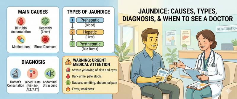
Yellowing of the skin or the whites of the eyes is a symptom difficult to ignore. Jaundice is not a disease — it is a signal: something has gone wrong with the processing or excretion of bilirubin. Behind this signal can lie entirely different conditions — from the benign Gilbert's syndrome to pancreatic cancer. This is precisely why the most important thing in jaundice is not the colour of the skin, but understanding the mechanism.
What Jaundice Is and Why It Occurs
Jaundice (icterus) is the yellowing of the skin, sclera, and mucous membranes due to the accumulation of bilirubin in the tissues. Visible jaundice appears when total bilirubin exceeds 34–40 µmol/L. At levels of 17–34 µmol/L, subclinical hyperbilirubinaemia exists — detectable on blood tests but invisible to the eye.
The most sensitive clinical indicator is the sclera: they contain abundant elastin, which binds bilirubin avidly. Scleral icterus appears before skin yellowing and persists longer during recovery.
Understanding the cause of jaundice comes down to one question: at which stage of bilirubin metabolism has the disruption occurred? Bilirubin is produced from haemoglobin breakdown, binds to albumin (indirect bilirubin), enters the liver, undergoes conjugation (direct bilirubin), and is excreted in bile. A breakdown at any of these stages produces a different type of jaundice.
Three Types of Jaundice: Mechanism and Distinctions
Prehepatic (haemolytic) jaundice
Mechanism: accelerated red cell destruction → more indirect bilirubin is produced than the liver can process.
Characteristic features:
- Mild to moderate yellowing (lemon-yellow hue)
- Dark urine from urobilinogen — not from direct bilirubin
- Dark stools — excess stercobilin
- Haemoglobin reduced; reticulocytes elevated
- Bilirubin: indirect elevated, direct — normal
- ALT, AST — normal or mildly elevated
Causes: haemolytic anaemia, malaria, incompatible blood transfusions, mechanical red cell destruction (prosthetic heart valves), Gilbert's syndrome.
Hepatic (parenchymal) jaundice
Mechanism: hepatocyte damage → simultaneous impairment of bilirubin uptake, conjugation, and excretion.
Characteristic features:
- Moderate to marked yellowing (saffron-yellow hue)
- Dark urine (direct bilirubin excreted by kidneys)
- Moderately pale stools
- ALT and AST markedly elevated (hepatocyte cytolysis)
- Both direct and indirect bilirubin elevated
- Albumin reduced; INR prolonged in severe disease
Causes: viral hepatitis A, B, C, E; alcoholic hepatitis; drug-induced hepatitis; liver cirrhosis; autoimmune hepatitis; non-alcoholic fatty liver disease.
Posthepatic (obstructive, cholestatic) jaundice
Mechanism: impaired bile outflow due to mechanical biliary obstruction → direct bilirubin cannot reach the intestine and accumulates in the blood.
Characteristic features:
- Marked yellowing (greenish hue with prolonged obstruction)
- Acholic (clay-coloured) stools — bile does not reach the intestine; stercobilin is not produced
- Dark "beer-coloured" urine — direct bilirubin excreted by the kidneys
- Skin itch — bile acids irritate cutaneous receptors
- ALP and GGT markedly elevated; ALT/AST moderately elevated
- Direct bilirubin sharply elevated; indirect — mildly elevated
Causes: gallstone disease with choledocholithiasis; pancreatic head cancer; cholangiocarcinoma; biliary strictures; primary sclerosing cholangitis.
Differential Table: Three Types of Jaundice
| Feature | Prehepatic | Hepatic | Posthepatic |
|---|---|---|---|
| Skin hue | Lemon-yellow | Saffron-yellow | Greenish-yellow |
| Urine colour | Dark | Dark | Dark ("beer") |
| Stool colour | Dark | Moderately pale | White (acholic) |
| Skin itch | Absent | Mild | Marked |
| ALT/AST | Normal | Markedly ↑ | Mildly ↑ |
| ALP/GGT | Normal | Mildly ↑ | Markedly ↑ |
| Direct bilirubin | Normal | ↑↑ | ↑↑↑ |
| Indirect bilirubin | ↑↑ | ↑ | Normal/↑ |
| Haemoglobin | ↓ | Normal | Normal |
Neonatal Jaundice: A Special Case
Jaundice in newborns is physiologically normal in the first days of life. After birth, fetal haemoglobin breaks down rapidly while conjugation enzymes are still immature. Peak physiological jaundice occurs on days 3–5 and resolves by days 7–10 in term infants.
When neonatal jaundice is pathological:
- Appears within the first 24 hours of life — always pathological
- Bilirubin exceeds age-specific thresholds (assessed using hour-specific nomograms)
- Persists beyond 2–3 weeks in term infants
- Direct bilirubin is elevated (sign of liver pathology or biliary atresia)
Critical elevation of indirect bilirubin in newborns → kernicterus (bilirubin encephalopathy): irreversible brainstem nuclei damage. Requires emergency phototherapy or exchange transfusion.
Diagnosing Jaundice: Where to Start
First step — blood tests:
- Bilirubin — total, direct, indirect: immediately identifies the jaundice type
- Liver function panel: ALT, AST, ALP, GGT, albumin — characterises the pattern of liver injury
- Complete blood count with reticulocytes — rules out haemolysis
- INR / prothrombin time — hepatic synthetic function
- C-reactive protein — inflammatory component
Second step — imaging: Abdominal ultrasound — mandatory for any jaundice beyond mild. Dilated bile ducts confirm mechanical obstruction. When tumour is suspected — CT or MRI with cholangiopancreatography (MRCP).
Third step — targeted tests based on the suspected cause:
- Viral hepatitis markers (HAV IgM, HBsAg, HCV RNA)
- Autoimmune markers (ANA, AMA, SMA)
- Direct biliary visualisation (ERCP) for mechanical obstruction
Treatment of Jaundice
Jaundice is a symptom, not a disease. Treatment is directed at the cause, not the skin colour.
- Haemolytic jaundice: treat the underlying disease — immunosuppression for autoimmune haemolytic anaemia, splenectomy for certain haemolytic conditions
- Hepatic jaundice: direct-acting antivirals for hepatitis C (cure > 95%); immunosuppression for autoimmune hepatitis; immediate alcohol cessation for alcoholic hepatitis; discontinue the offending drug in drug-induced hepatitis
- Obstructive jaundice: endoscopic stone removal (ERCP), biliary stenting for malignant obstruction, surgical treatment
In obstructive jaundice, biliary decompression is critical: prolonged cholestasis damages hepatocytes and leads to secondary biliary cirrhosis.
When to Seek Urgent Medical Attention
Call emergency services immediately if jaundice is accompanied by:
- Fever, chills, and right upper quadrant pain — Charcot's triad: cholangitis, a life-threatening biliary infection
- Confusion — hepatic encephalopathy in decompensated cirrhosis
- Severe abdominal pain — possible perforation or acute pancreatitis
- Rapidly worsening jaundice over a few days
Schedule a routine appointment for:
- Any newly appeared jaundice or scleral icterus in an adult
- Acholic stools even with mild jaundice — suspicion of obstruction
- Jaundice in a pregnant woman — obstetric cholestasis requires immediate assessment
- Jaundice in a newborn within 24 hours of birth or persisting beyond 2 weeks
This article is for informational purposes only and does not replace consultation with a gastroenterologist or GP.
For informational purposes only
This article is for informational purposes only and does not constitute medical advice, diagnosis, or treatment. Please consult a healthcare professional for medical guidance.