Varicocele: Symptoms, Grades and Scrotal Ultrasound Doppler
Reviewed by the LabReadAI medical team
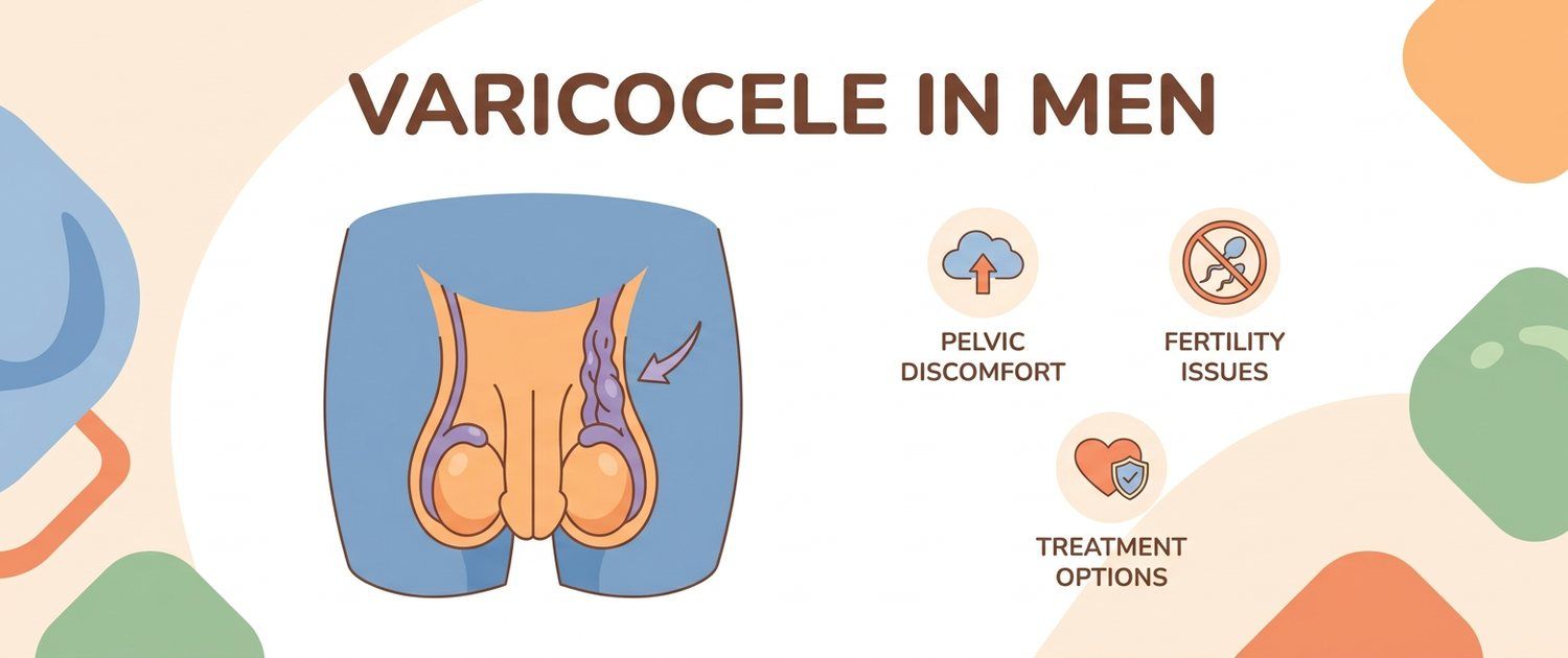
Varicocele is one of the most common male findings on exam and scrotal ultrasound, and at the same time one of the most alarming to hear. In fact it is an enlargement of the veins around the testicle that often causes no complaints and is found by chance. Let's look at what symptoms occur, why the left side is affected more often, what grades I–III mean and how varicocele relates to fertility.
What Varicocele Is
Varicocele is the enlargement and tortuosity of the veins of the so-called pampiniform plexus of the scrotum, similar to varicose veins in the legs but in a different place. Blood drains through the veins less well, they overfill and enlarge. The enlargement itself is not a tumor or an infection but a vascular feature that in some men causes no problems at all.
Why Varicocele Is More Often on the Left
Varicocele more often occurs on the left because of anatomy: the left testicular vein is longer and drains into the renal vein at a right angle, which makes outflow harder. Right-sided and bilateral varicocele also occur but are rarer. An isolated, rapidly appearing right-sided enlargement of the veins is a reason for a more careful exam by a doctor.
Varicocele Symptoms
Varicocele is often symptomless and found by chance. When symptoms are present, they are usually heaviness and a dragging or aching pain in the scrotum that worsens toward evening, with exertion and in heat, and eases when lying down. Sometimes a man feels a "bag of worms" or notices that one side of the scrotum is slightly larger. Sudden severe pain is not typical varicocele and is a reason to see a doctor urgently.
Varicocele Grades I–III
The grades describe how pronounced the enlarged veins are on exam: I — veins are felt only on straining (Valsalva maneuver), II — felt at rest, III — visible through the scrotal skin. Separately, a subclinical varicocele is defined, which is not felt and is seen only on ultrasound. The grade alone does not decide management — it is assessed together with symptoms and semen values.
Scrotal Ultrasound with Doppler
The main confirming method is a scrotal ultrasound with color Doppler. It shows the diameter of the enlarged veins and reverse blood flow (reflux) on straining, as well as the state of the testicles themselves. Doppler helps distinguish a true varicocele from an incidental finding and assess subclinical forms. If the ultrasound report is unclear, you can upload the scan for decoding — the service explains the wording in plain language.
Varicocele and Male Infertility
Varicocele is a common reversible factor affecting fertility: overheated enlarged veins can worsen a spermogram — lowering concentration, motility and the share of normal forms. Yet many men with varicocele have a normal spermogram and father children without problems. So the link is judged not by the presence of varicocele but by tests and family plans within an infertility work-up.
When to See a Doctor
See a urologist for pain and heaviness in the scrotum, a noticeable "bag of veins", a difference in testicle size, and also when planning children and with abnormal semen values. Separately, the prostate is checked — for example, with complaints a prostate ultrasound (TRUS) helps. Whether surgery is needed and when is decided by a doctor from all the data, not from one ultrasound.
This article is for informational purposes only and does not replace a doctor's consultation. The grade of varicocele and management are assessed by a specialist from the exam, ultrasound and spermogram.
For informational purposes only
This article is for informational purposes only and does not constitute medical advice, diagnosis, or treatment. Please consult a healthcare professional for medical guidance.