AMH (Anti-Müllerian Hormone): Normal Levels by Age and Reserve
Reviewed by the LabReadAI medical team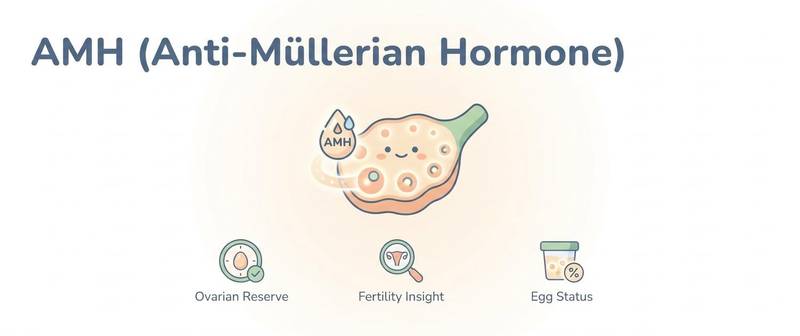
If FSH shows how the body is reacting to the ovaries "right now", anti-Müllerian hormone answers a more strategic question: how much "reserve" is left. AMH is produced by small growing follicles, and its level directly reflects ovarian reserve — the pool of eggs available going forward. This makes AMH a key marker for pregnancy planning and IVF preparation. Here is how to read it.
What AMH Is and What It Shows
Anti-Müllerian hormone (AMH) in women is produced by granulosa cells of pre-antral and small antral ovarian follicles. The larger this follicle pool, the higher the AMH — which is why it serves as a marker of ovarian reserve. Unlike FSH and oestradiol, whose levels swing across the cycle, AMH is stable — its main practical advantage.
Men also have AMH (produced by Sertoli cells), used in paediatric endocrinology, but in adult practice it is primarily a "female" marker of ovarian reserve.
AMH Normal Levels by Age
AMH declines steadily with age as the follicle pool naturally depletes:
| Age | Approximate normal AMH, ng/mL |
|---|---|
| 20–29 years | 1.5–6.8 |
| 30–34 years | 1.1–5.0 |
| 35–39 years | 0.7–3.5 |
| 40–44 years | 0.3–2.0 |
| After 45 years | often < 0.5 |
Reference ranges depend on the laboratory assay — always check your report. Units may be ng/mL or pmol/L (1 ng/mL ≈ 7.14 pmol/L).
The decline of AMH with age is normal, not a disease. What matters clinically is not the number itself but how it fits the age and the goal (for example, pregnancy planning).
When to Test AMH in the Cycle and How to Prepare
The main convenience of AMH is that it can be tested on any cycle day, because it barely fluctuates across phases. Blood is drawn from a vein; strict fasting is not required, though a morning sample is standard. On combined oral contraceptives the AMH level may be somewhat reduced — this is taken into account when interpreting the result.
Low AMH: Causes and What It Means for Fertility
Low AMH indicates diminished ovarian reserve — few follicles remain. Causes:
- age — the main, natural factor;
- premature decline / premature ovarian insufficiency (together with high FSH);
- ovarian surgery, chemo- or radiotherapy;
- smoking, endometriosis.
It is important to understand: low AMH reflects egg quantity, not quality, and does not mean infertility. Many women with low AMH conceive naturally. But when planning a pregnancy, low AMH is a reason not to delay and to discuss the approach with a reproductive specialist. A sharp drop at a young age is interpreted together with signs of menopause and the FSH level.
High AMH: PCOS and Other Causes
Elevated AMH is most often linked to a large number of small follicles — typical of polycystic ovary syndrome. In PCOS, high AMH reflects an excess of antral follicles and correlates with the severity of ovulation problems. Less commonly, high AMH occurs with certain ovarian tumours. A very high level also matters before IVF — it warns of the risk of hyperstimulation syndrome.
AMH, IVF and Ovarian Reserve Assessment
In IVF programmes AMH is one of the main parameters: it helps predict the ovarian response to stimulation and choose the drug dose.
- Very low AMH → a "poor response" is expected, few eggs; the strategy is individualised.
- Very high AMH → risk of hyperstimulation; the dose is reduced.
But AMH is never assessed in isolation. The full reserve picture is AMH + basal FSH + antral follicle count on ultrasound, and planning also considers other hormones from the sex hormone panel.
AMH Interpretation: With FSH and Ultrasound, and When to See a Doctor
- Low AMH + high FSH in a young woman — a sign of diminished reserve; a reproductive specialist is needed, especially when planning a pregnancy.
- High AMH + cycle disturbances + signs of androgen excess — a pattern consistent with PCOS; the diagnosis is made on the whole picture.
- Borderline AMH — assessed over time and in the context of age, without hasty conclusions from a single number.
For a detailed breakdown of the female hormone profile by age, see the article which hormone tests women should get after 40.
This article is for informational purposes. Interpretation of the test and the management plan are the job of a gynaecologist or reproductive specialist.
For informational purposes only
This article is for informational purposes only and does not constitute medical advice, diagnosis, or treatment. Please consult a healthcare professional for medical guidance.