Urinalysis Interpretation: How to Read Every Result
Reviewed by the LabReadAI medical team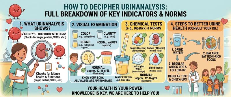
You are holding a results sheet with two dozen rows — colour, pH, protein, leukocytes, casts, nitrites — and do not know where to start. Urinalysis is one of the most commonly ordered laboratory tests, and at the same time one of the least understood by patients. This article walks through every indicator in order: what is being measured, what value counts as normal, what deviations mean, and how serious they are. Not to replace your doctor — but so you can arrive at the appointment prepared and understand what is being discussed.
How a Urinalysis Is Structured and How to Read It
A urinalysis consists of three sequential components:
- Physical properties — what can be seen with the naked eye and assessed with a urinometer: colour, clarity, odour, specific gravity, and acidity (pH)
- Chemical analysis — a dipstick detects the presence or absence of substances that should not normally appear in urine: protein, glucose, ketones, bilirubin, nitrites
- Sediment microscopy — after centrifuging the sample, the laboratory technician examines the sediment under a microscope, counting cells, casts, and crystals
The three components complement each other. Physical properties give an overall first impression. Chemistry clarifies: are there pathological substances present? Microscopy shows where they come from — glomeruli, tubules, bladder, or prostate.
How to read the report: next to each parameter, the laboratory prints its reference range — the normal value for its specific method. Compare against this, not against averaged internet figures: norms differ between equipment and reagents.
Physical Properties: Colour, Clarity, Specific Gravity and pH
Colour — the first thing the technician evaluates. Normal: straw-yellow to amber. Intensity depends on hydration: a dehydrated body produces dark concentrated urine; a well-hydrated one produces light urine. Deviations:
- Red or pink — blood (haematuria), beetroot, rifampicin
- Dark brown, "strong tea" colour — bilirubinuria, liver disease
- Cloudy white — pyuria in severe infection, phosphaturia
- Nearly colourless — polyuria, diabetes insipidus, excessive fluid intake
Clarity — normally full. Turbidity indicates bacteria, leukocytes, mucus, epithelium, or salts. Mild turbidity from a sample left standing is an artefact, not pathology.
Specific gravity — normal 1.010–1.025. Reflects the kidneys' ability to concentrate urine. Persistently low density (hyposthenuria < 1.010) indicates impaired concentrating function, characteristic of chronic renal insufficiency. High density (> 1.030) — dehydration, diabetes mellitus.
pH — normal 4.5–8.0. Acidic urine is typical of a high-meat diet, fever, and gout. Alkaline urine — vegetarian diet, urinary tract infection (bacteria break down urea into ammonia), prolonged sample storage.
Chemical Indicators: Protein, Glucose and Ketones
Protein (proteinuria) — normal: absent or trace up to 0.033 g/L. Protein in urine is one of the most important signals of kidney filter damage. Even a small excess warrants attention.
Types and causes of proteinuria:
- Physiological (functional) — after strenuous exercise, fever, or cold exposure. Disappears at rest.
- Orthostatic — in young people, protein appears only in daytime upright urine, absent at night. A benign variant.
- Pathological glomerular — in glomerulonephritis, nephrotic syndrome, diabetic nephropathy. Protein > 1–3 g/L, often combined with red blood cells and casts.
- Tubular — from tubular damage: tubulointerstitial nephritis, Fanconi syndrome.
For a detailed article on causes and clinical significance, see protein in urine.
Glucose (glycosuria) — normal: absent. Appears when blood glucose exceeds the renal threshold (~9–10 mmol/L). Main causes: diabetes mellitus, stress hyperglycaemia, renal glycosuria (impaired tubular reabsorption with normal blood sugar). Glucose in urine without hyperglycaemia is a reason to check tubular function.
Ketone bodies — normal: absent. Acetone, acetoacetate, and beta-hydroxybutyrate appear with accelerated fat breakdown: fasting, low-carbohydrate diet, diabetic ketoacidosis, alcoholic ketoacidosis, severe vomiting in pregnancy. A distinction is important: trace ketones on a ketogenic diet are an expected finding. Marked ketonuria with symptoms is an emergency. Everything about ketones — in ketones in urine.
Liver Function Markers: Bilirubin and Urobilinogen
Bilirubin — normal: absent. Only direct (conjugated) bilirubin, being water-soluble, passes through the kidney filter. Its presence in urine signals cholestasis or parenchymal liver disease: viral hepatitis, cirrhosis, gallstone disease with obstruction, pancreatic cancer. Urine darkens and froths when shaken.
Indirect (unconjugated) bilirubin does not enter urine — it is bound to albumin and water-insoluble. This is why in haemolytic anaemia with elevated indirect bilirubin, urine remains light-coloured.
Urobilinogen — a product of bilirubin reduction by intestinal bacteria. Normal: up to 17 µmol/L. Elevated in haemolysis (increased red cell breakdown), impaired hepatic excretion (hepatitis, cirrhosis), and constipation. Complete absence of urobilinogen alongside bilirubinuria indicates total bile duct obstruction.
Nitrites and Bacteria: Infection Markers
Nitrites — normal: absent. Gram-negative bacteria (E. coli, Klebsiella, Proteus) convert urinary nitrates to nitrites. A positive nitrite test from a properly collected sample is a highly specific sign of bacterial infection.
An important limitation: gram-positive bacteria (staphylococci, enterococci) do not possess nitrate reductase — when infection is caused by them, nitrites will be negative even with a high bacterial count. A negative nitrite result does not rule out infection.
Bacteria — normally absent in a properly collected sample. Bacteria on microscopy combined with pyuria is grounds for ordering a urine culture with antibiotic sensitivity. Bacteria without pyuria suggests sample contamination.
Sediment Microscopy: Leukocytes, Red Blood Cells and Casts
Leukocytes — normal: up to 5 per high-power field in women, up to 3 in men. Pyuria (leukocyturia) means inflammation in the urinary tract. The level roughly reflects severity: 5–15 mild, 15–50 moderate, above 50 severe. A full breakdown of the indicator: leukocytes in urine.
Key for interpretation: pyuria without nitrites or bacteria is called sterile pyuria. Causes include renal tuberculosis, chlamydial infection, analgesic nephropathy, and interstitial nephritis — situations where a standard urine culture is negative but inflammation is present.
Red blood cells — normal: 0–3 per high-power field. Haematuria — blood in urine. The character of red cells suggests the source:
- Intact (fresh) red blood cells — bleeding from the lower tract: bladder, urethra, prostate. Causes: cystitis, kidney stone, tumour.
- Dysmorphic ("ghost") red blood cells — have passed through the glomerular filter. Sign of glomerulonephritis. Their presence with protein and casts = nephritic syndrome.
Casts are protein moulds formed in renal tubules. Their presence indicates pathology specifically in the renal parenchyma.
| Cast type | Composition | Significance |
|---|---|---|
| Hyaline | Pure protein | Occasional — normal; many — proteinuria, exercise |
| Granular | Protein + cellular debris | Tubular damage, chronic disease |
| Waxy | Dense protein | Chronic renal failure, amyloidosis |
| Red blood cell | Protein + RBCs | Glomerulonephritis, vasculitis — pathognomonic |
| White blood cell | Protein + WBCs | Pyelonephritis, interstitial nephritis |
| Epithelial | Protein + tubular epithelium | Acute tubular necrosis, nephrotoxins |
Red blood cell and waxy casts are alarming findings requiring urgent nephrology consultation.
Epithelial cells — occasional transitional and squamous cells are acceptable. Massive shedding of renal (tubular) epithelium indicates acute tubular necrosis or nephrotoxic injury.
Crystals — precipitated salts. Occasional oxalate or urate crystals at normal urine output are not pathological. Massive crystalluria with flank pain suggests nephrolithiasis. Cystine crystals are a rare marker of cystinuria.
Mucus — trace amounts are acceptable. Abundant mucus indicates sample contamination or chronic urethral irritation.
How to Read a Urinalysis Report: A Step-by-Step Approach
A few practical steps for independent orientation:
Start with the laboratory's reference range — printed next to each parameter. This, not internet averages, is your baseline.
Distinguish isolated from combined abnormalities. A single isolated finding (trace protein with no leukocytes or casts) is generally less significant than a combination (protein + red blood cell casts + high leukocytes).
Match the result to symptoms. Pyuria with burning on urination and positive nitrites is most likely cystitis. The same pyuria without symptoms in an elderly man is a different diagnostic context entirely.
Check collection conditions. A contaminated sample (abundant squamous epithelium, heavy mucus) makes the entire test unreliable. If in doubt — repeat with proper technique.
Do not diagnose from a single test. A one-off abnormality warrants a repeat. Consistent abnormalities on two or more tests are the basis for further investigation.
When Urinalysis Results Require Urgent Medical Attention
Most findings call for a scheduled appointment. But certain results demand immediate action:
- Visible blood in urine or red blood cells > 50 per field — urgent
- Red blood cell or waxy casts — nephrology emergency
- Protein > 1 g/L combined with oedema and elevated blood pressure — nephrotic or nephritic syndrome
- Pyuria > 50 per field + fever + flank pain — possible pyelonephritis
- Marked glycosuria with ketones and symptoms — possible diabetic ketoacidosis
- Any pathological finding in a pregnant woman — immediate consultation
This content is for informational purposes only and does not replace professional medical advice.
For informational purposes only
This article is for informational purposes only and does not constitute medical advice, diagnosis, or treatment. Please consult a healthcare professional for medical guidance.