Cat Watery Eye: Causes, Corneal Ulcers and Red Flags
Reviewed by the LabReadAI medical team
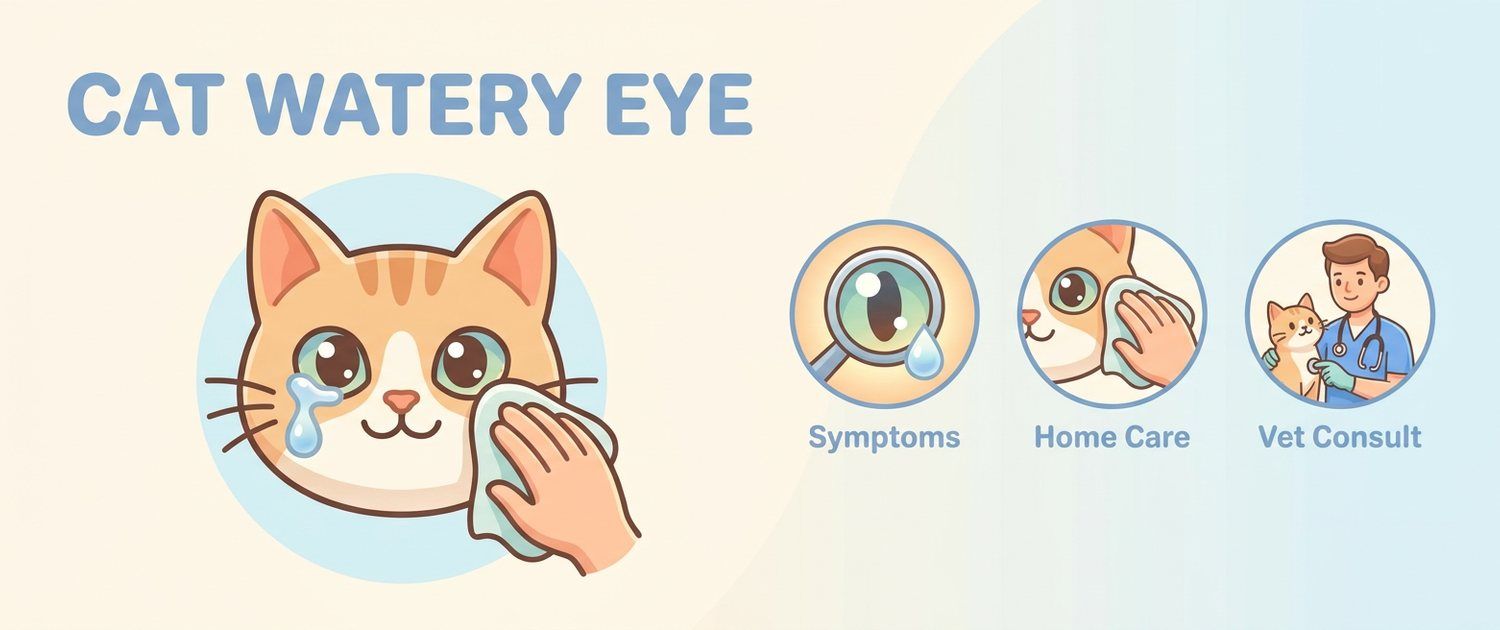
A watery eye looks the same in almost every case, from a speck of dust under the lid to an ulcer that ends with the eye being removed. So the point of this article is not to guess a diagnosis from a photo but to answer one question correctly: can this wait until tomorrow, or does it go to the clinic now.
Cat Watery Eye Causes, Ranked by Frequency
- Conjunctivitis — inflammation of the lid lining. The commonest and usually the least dangerous answer.
- Herpesvirus infection — in cats the leading cause of chronic and recurrent watering.
- Trauma and foreign bodies — a grass blade, a claw from a fight, a grain of sand. Almost always one eye.
- Corneal ulcer — the dangerous one, and it often follows the first two.
- Impaired tear drainage — narrow or blocked ducts, an inturned eyelid.
- Uveitis — inflammation inside the eye from systemic disease: toxoplasmosis, feline infectious peritonitis, leukaemia, lymphoma.
- Glaucoma and tumours — rare, but they need to be recognised fast.
Cat Conjunctivitis: Common and Usually Benign
With conjunctivitis the eye reddens and waters but stays open: the cat neither hides it nor avoids light. Discharge starts clear and may thicken later. Both eyes are often involved, because an infection is usually behind it.
The key idea: conjunctivitis is the default assumption, but it cannot be assigned without excluding an ulcer. In the first hours they look alike, and their outcomes do not.
Corneal Ulcer in Cats: Squinting Means Hours, Not Days
The cornea is the clear front wall of the eye. Damage to it hurts enough that the cat screws the eye shut, turns away from light and hides. The picture then moves fast: clouding, a bluish or white area, sometimes a visible dent in the surface.
Why missing an ulcer costs the eye:
- a deep ulcer can perforate within 24–48 hours;
- after perforation the inner structures prolapse and the eye is usually lost;
- the classic owner error is reaching for leftover drops. Steroid drops applied to an ulcer accelerate its deepening, and they feature in most stories of eyes that were lost.
A vet finds an ulcer in a minute: fluorescein stain makes the defect visible. That cannot be done at home, which is why a squinting eye always travels to the clinic.
Herpesvirus and Recurrent Watering
Feline herpesvirus attacks both the conjunctiva and the cornea, and after recovery it retreats into nerve ganglia and stays for life. Hence the familiar story: "the eye healed six months ago, and now it is back after we moved house." That is a flare, not a fresh infection.
A hallmark of the virus is branching, dendritic corneal ulcers, which are close to specific for it. In kittens it usually travels with sneezing and a blocked nose, so the eye is only part of the picture. If mouth ulcers and drooling appear too, that leans towards calicivirus — see drooling in cats.
One Eye Watering in a Cat: What That Changes
One-sidedness cuts the list sharply:
| Both eyes | One eye |
|---|---|
| Viral or bacterial infection | Trauma, a scratch |
| Allergy and irritants | Foreign body under the third eyelid |
| Systemic disease | Entropion, blocked tear duct |
| Smoke, dust, draught | Ulcer, uveitis, rarely a tumour |
The practical conclusion: a one-sided watery eye needs an exam sooner, even in a cat that seems comfortable. Allergy in cats, incidentally, shows up on the skin far more often than in the eyes — see a scratching cat.
Cat Eye Discharge: What Its Character Means
- Clear and watery — irritation, an early viral phase, or poor tear drainage.
- Mucoid and stringy — a drawn-out inflammation.
- Thick yellow-green, lids gummed shut in the morning — bacteria have joined in.
- Rusty brown streaks below the eye — not blood but oxidised tear pigment, a sign that tears run down the face instead of into the duct.
Colour is useful, but it does not answer the main question of whether the cornea is damaged. Only an exam does.
Flat-Faced Breeds and Kittens: Two Separate Cases
In Persians, Exotics and other flat-faced cats the tears run over because they have nowhere to drain: the puncta and ducts are distorted by the skull, and the lids do not close perfectly. That watering is chronic, bilateral and painless; it calls for grooming rather than alarm — but any pain or clouding on top of it reads as a new problem, not as "the breed".
In a kitten a watery, gummed-shut eye is nearly always infection, and it is dangerous because the lids can adhere to the cornea. In very young kittens pus can build up behind lids that have not yet opened. If the kitten also stops eating, follow the logic in why a cat is not eating.
When It Is Urgent: Cat Red Eye and Other Red Flags
Go today rather than watch if any of these are present:
- the cat squints, keeps the eye shut, avoids light;
- a cloudy, white or bluish patch on the cornea, or a visible dent;
- pupils of different sizes, or an eye that looks enlarged or glassy;
- blood inside the eye, prolapsing tissue, a torn eyelid;
- sharp deterioration over a few hours;
- the eye plus systemic signs — lethargy, fever, refusing food; see a lethargic cat;
- watering in a cat that is losing weight, which points to a systemic cause; the reasoning is in a cat losing weight.
What Not to Do at Home
- No human medicines. Human painkillers and fever drugs are lethal to cats. Any medication, eye drops included, is a vet's decision only.
- No steroid drops or ointments "like last time": with an ulcer present they speed up its deepening.
- No tea, chamomile, boric acid or alcohol-based rinses.
- Do not fish out a foreign body with tweezers or dry cotton.
- Do not wait out the weekend if the cat is squinting: a day matters here.
- Do not cover or tape the eye.
What is safe: wipe crusts off the lids with clean gauze moistened with sterile saline, working from the nose outwards, and stop the cat rubbing its face — a collar fitted by the vet does that better than any home improvisation.
What Tests and Imaging Will Clarify the Causes
Eyes are examined mostly with instruments rather than blood work, but recurrent and systemic pictures need the laboratory too:
- Fluorescein stain — is there a corneal defect. The basic and most important step.
- Schirmer tear test — is tear production sufficient; dry eye is uncommon in cats but real.
- Intraocular pressure — glaucoma versus uveitis.
- PCR for herpesvirus and chlamydia from swabs — for chronic, relapsing disease.
- Blood count, biochemistry and tests for feline leukaemia and immunodeficiency virus — when uveitis is suspected; how to read the form is in cat blood test.
- Ocular ultrasound and orbital tomography — for a bulging eye, trauma or a suspected mass behind the globe; see MRI and CT in dogs and cats, while tooth roots and sinuses are assessed on a cat X-ray.
Other species read differently: in rabbits a watering eye is most often driven by tooth roots pressing on the tear duct — see diseases of rabbits, tortoises and rodents.
A lab form from the clinic can be read in the pet results interpretation service, a scan or report in pet imaging interpretation, and if nothing has been tested yet you can describe the situation to get the likely causes and the urgency. How a document-based read works is explained in online vet reading.
This article is informational. Diagnosis and treatment of your pet are the job of a veterinarian.
For informational purposes only
This article is for informational purposes only and does not constitute medical advice, diagnosis, or treatment. Please consult a healthcare professional for medical guidance.