Calcitonin Blood Test: Normal Levels, Results and Thyroid Cancer
Reviewed by the LabReadAI medical team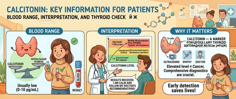
You had thyroid hormone tests done — and the report includes calcitonin with an upward arrow. Or your doctor ordered it before a planned thyroid procedure. Either way the same question arises: what does it mean? Calcitonin is one of the few hormones that simultaneously regulates calcium metabolism and serves as a highly specific tumour marker. This article explains what it measures, what the reference ranges are, and why an elevated result must not be overlooked.
What Is Calcitonin and What Does It Do
Calcitonin is produced by parafollicular C-cells of the thyroid gland — a small population of cells scattered among the follicular thyrocytes. Unlike thyroxine and triiodothyronine, calcitonin does not depend on TSH: it is regulated directly by blood calcium levels. When calcium rises, calcitonin secretion increases in response.
Physiologically, calcitonin lowers blood calcium by inhibiting osteoclast-mediated bone resorption and increasing urinary calcium excretion. In clinical practice this function has limited importance — even complete calcitonin deficiency after total thyroidectomy does not cause meaningful disturbance of calcium metabolism in adults.
The main reason calcitonin is measured clinically is entirely different: thyroid C-cells are the origin of medullary thyroid carcinoma, and the tumour retains the ability to secrete calcitonin in large quantities. This makes calcitonin a sensitive and specific tumour marker — one of the few in endocrinology with genuine diagnostic value.
The test is ordered for: thyroid nodules before biopsy or surgery; suspected medullary thyroid cancer on ultrasound or cytology; hereditary MEN-2 syndromes (multiple endocrine neoplasia); post-operative surveillance after medullary thyroid cancer surgery; and family history of medullary thyroid cancer or phaeochromocytoma.
How to Prepare for a Calcitonin Test
Blood is drawn from a vein, preferably in the morning. Calcitonin levels are sensitive to several physiological factors — ignoring preparation requirements can produce falsely elevated results and unnecessary anxiety.
Fasting: strictly required — no food for at least 8–12 hours. Even a light meal stimulates calcitonin secretion via a gastrin-dependent mechanism and can raise the result 1.5–2 fold.
Smoking: nicotine stimulates calcitonin secretion. Refrain from smoking for at least 2 hours before the blood draw — ideally from the evening before.
Physical activity and stress: avoid intense exercise for 24 hours. Acute physical or emotional stress activates the sympathetic nervous system and can transiently elevate calcitonin.
Medications: proton pump inhibitors (omeprazole, pantoprazole), pentagastrin, and calcium supplements affect calcitonin levels. Discuss temporary discontinuation with your doctor before a planned test.
Stimulation test: in diagnostically challenging cases, an intravenous calcium or pentagastrin stimulation test is used. Stimulated calcitonin is far more sensitive than the basal value — particularly for early-stage medullary cancer and carriers of RET mutations. This test is performed only in specialist inpatient settings.
The assay uses immunochemiluminescence (ICLA) or electrochemiluminescence (ECLA). Results are ready within 1–2 business days.
Calcitonin Normal Range: Reference Values
Calcitonin reference ranges differ substantially between men and women — one of the few situations in laboratory diagnostics where sex is critically important. Different analyser generations and reagents also produce slightly different thresholds; always use the range on your own report.
| Group | Basal Calcitonin | Borderline Zone | Significantly Elevated |
|---|---|---|---|
| Men | < 9.52 pg/mL | 9.52–100 pg/mL | > 100 pg/mL |
| Women | < 6.4 pg/mL | 6.4–30 pg/mL | > 30 pg/mL |
| Infants under 6 months | up to 40 pg/mL | — | > 100 pg/mL |
The borderline zone deserves a separate note. A result in the range of 10–100 pg/mL in men or 6–30 pg/mL in women does not automatically indicate cancer: such values occur in C-cell hyperplasia (a benign condition), chronic kidney disease, and neuroendocrine tumours of other origins. A stimulation test and repeat measurement in follow-up are the standard approach in this zone.
Values above 100 pg/mL in an adult — regardless of sex — are considered highly suspicious for medullary thyroid carcinoma and require immediate oncological evaluation. At levels above 500 pg/mL the probability of medullary cancer approaches 100%.
After successful surgery for medullary thyroid cancer, calcitonin should fall to undetectable levels. Persistent elevation or renewed rise on follow-up measurement indicates residual disease, recurrence, or metastases.
Calcitonin as a Tumour Marker for Medullary Thyroid Cancer
Medullary thyroid carcinoma accounts for approximately 5–10% of all thyroid malignancies but follows a more aggressive course than papillary or follicular types. Around 25% of cases are linked to inherited RET gene mutations — these patients often present as part of MEN-2A or MEN-2B syndromes, in which medullary cancer is combined with phaeochromocytoma and hyperparathyroidism.
The key distinction of calcitonin from most tumour markers is its high specificity. PSA, for example, rises with any prostate inflammation and is not a strict marker of malignancy. Calcitonin, by contrast, rises almost exclusively in C-cell pathology — giving it a diagnostic precision that is rare among oncological markers.
In a comprehensive workup for suspected thyroid malignancy, calcitonin is assessed alongside CEA — carcinoembryonic antigen, also secreted by medullary cancer cells. Simultaneous elevation of both markers is a characteristic combination confirming the diagnosis. The ratio between the two carries prognostic weight: rapid CEA growth while calcitonin stabilises may indicate tumour dedifferentiation.
For a complete thyroid hormonal workup, calcitonin is included in the extended thyroid panel. The standard panel (TSH, T4, T3) does not include calcitonin — it is added specifically when nodular lesions are present.
In 2022 the European Society of Endocrinology recommended routine calcitonin measurement in all patients with thyroid nodules before biopsy or surgery decisions. This enables detection of medullary cancer at an early stage — when surgical outcomes are best.
Non-Oncological Causes of Elevated Calcitonin
Elevated calcitonin does not always mean cancer. The list of benign causes is substantial, and a clinician will always consider these before initiating extended oncological evaluation.
C-cell hyperplasia. A benign increase in C-cell numbers — often a precursor to medullary cancer in RET mutation carriers, but in most patients a stable finding. Calcitonin is mildly elevated, and the stimulation test may be positive. Requires dynamic monitoring.
Chronic kidney disease. The kidneys participate in calcitonin clearance. As glomerular filtration rate declines, the hormone accumulates — often reaching borderline values.
Neuroendocrine tumours of other sites. Certain lung, pancreatic, and intestinal carcinoids can ectopically secrete calcitonin. Levels are usually moderately elevated.
Acute conditions: severe sepsis, acute pancreatitis, and extensive burns produce reactive calcitonin elevation — sometimes reaching 100–200 pg/mL. This is an acute-phase response unrelated to oncology.
Autoimmune thyroiditis during flares can produce mild elevation, likely from inflammatory stimulation of C-cells.
Physiological factors: pregnancy and lactation are accompanied by expected calcitonin rises as part of calcium metabolism adaptation.
Low Calcitonin: When Is It a Problem
A result of "below normal" or "undetectable" is common — particularly in women, where the basal level may fall below the analyser detection threshold. This is not pathological.
After total thyroidectomy, calcitonin becomes undetectable: the gland is removed, C-cells are gone. This is the expected and desirable outcome when monitoring after medullary thyroid cancer surgery.
Clinically significant calcitonin deficiency is extremely rare and does not typically require treatment — unlike deficiency of TSH-regulated thyroid hormones.
When to See a Doctor
Prompt evaluation by an endocrinologist or oncologist is required when:
- Calcitonin exceeds 100 pg/mL in an adult under any conditions — the threshold for high oncological concern.
- Elevated calcitonin is found alongside a thyroid nodule on ultrasound — the combination warrants urgent biopsy and oncology consultation.
- The result exceeds 500 pg/mL — the probability of medullary cancer is critically high; hospitalisation and staging workup are needed.
- Calcitonin is rising on serial measurements — even if absolute values are moderate, the trend matters more than any single result.
- Family history includes medullary thyroid cancer, phaeochromocytoma, or MEN-2 syndrome — planned screening is mandatory even without symptoms.
- Calcitonin fails to fall to zero after thyroid surgery — a sign of incomplete tumour removal or early recurrence.
This article is for informational purposes only and does not replace medical consultation. Result interpretation and diagnosis are made by a qualified physician.
For informational purposes only
This article is for informational purposes only and does not constitute medical advice, diagnosis, or treatment. Please consult a healthcare professional for medical guidance.