CEA Tumour Marker: Normal Levels, Causes and Interpretation
Reviewed by the LabReadAI medical team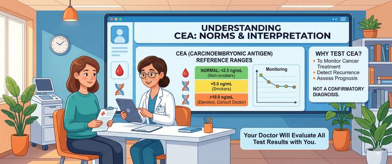
A blood test shows elevated CEA — and the first thought is cancer. That fear is understandable but rarely justified. Carcinoembryonic antigen rises in dozens of conditions that have nothing to do with malignancy, and is not a diagnostic test for cancer in the first place. Here is what CEA actually measures, when it genuinely matters clinically, and how to make sense of the result.
What Is CEA and Why Is It Ordered
CEA (carcinoembryonic antigen) is a glycoprotein produced in large amounts by the fetal gastrointestinal tract during intrauterine development. After birth, synthesis drops sharply, and in healthy adults CEA is detectable in the blood only at trace concentrations.
The most important thing to understand immediately: CEA is not a test to diagnose cancer. It can neither confirm nor rule out a malignant tumour in someone without an established diagnosis. CEA's sensitivity and specificity are insufficient for population screening — many patients with early-stage cancer have normal levels, and many healthy people are mildly elevated.
CEA's real clinical value lies in monitoring:
- Assessing treatment response — following chemotherapy, radiotherapy, or surgery. A falling CEA after treatment indicates effectiveness.
- Early detection of recurrence — in patients with treated colorectal cancer, regular CEA monitoring can detect disease return 3–6 months before clinical symptoms or CT changes appear.
- Staging — very high CEA at initial diagnosis correlates with metastatic disease and carries a worse prognosis.
The test is part of the standard tumour marker panel and is ordered for specific clinical indications, never as a preventive screen.
CEA Normal Range: Values for Different Groups
Reference ranges depend on the assay method and laboratory. The values below are approximate.
| Group | Normal range |
|---|---|
| Non-smoking adults | < 3.0 ng/mL (µg/L) |
| Smoking adults | < 5.0 ng/mL (µg/L) |
Why is the threshold higher for smokers? Tobacco smoke directly stimulates CEA synthesis in the bronchial and gastrointestinal mucosa. This is a physiological response to chronic irritation, not a sign of disease. Former smokers typically return to non-smoking reference values within several months of quitting.
Interpretation zones (approximate, for non-smokers):
- < 3.0 ng/mL — normal; oncological pathology is an unlikely sole explanation
- 3–10 ng/mL — mild elevation; requires clinical context. Non-oncological causes are more common at this range
- > 10 ng/mL — significant elevation; probability of malignancy is higher; detailed investigation needed
- > 20 ng/mL — high probability of advanced malignancy, especially when symptoms are present
In monitoring: the absolute value matters far less than trend. A drop from 50 to 8 ng/mL is a positive treatment response, even if 8 ng/mL exceeds the reference limit.
Which Cancers Are Associated with Elevated CEA
CEA is a non-specific tumour marker — it may rise in several types of malignancy.
Colorectal cancer — the primary clinical application for CEA. The marker is elevated in 70–80% of patients with metastatic colorectal cancer and in 40–50% with localised disease. After curative resection, CEA should normalise within 4–8 weeks. Failure to do so suggests residual metastases. Post-operative CEA monitoring for colorectal cancer is typically performed every 3 months for the first 2–3 years.
Lung cancer, especially adenocarcinoma — CEA is elevated in approximately 40–60% of patients and is used as a supplementary marker during treatment monitoring.
Breast, gastric, pancreatic, medullary thyroid and ovarian cancers — CEA may be mildly elevated in these malignancies but is not the marker of choice.
A fundamental limitation: CEA is normal in early-stage disease for most tumour types. A negative result does not exclude cancer.
Non-Oncological Causes of Elevated CEA
This is the most underappreciated category. The majority of mild CEA elevations (3–10 ng/mL) are explained by conditions unrelated to cancer.
Liver disease:
- Liver cirrhosis — impaired CEA clearance by damaged hepatocytes; chronically elevated CEA in cirrhosis does not mean cancer
- Chronic hepatitis — particularly viral; CEA is mildly elevated during active inflammation
- Alcoholic liver disease
Inflammatory bowel disease — Crohn's disease and ulcerative colitis produce a mild chronic CEA elevation from mucosal inflammation.
Gallstone disease and pancreatitis — biliary obstruction from any cause impairs CEA clearance, causing it to accumulate.
Benign pulmonary conditions — COPD, bronchiectasis, chronic pneumonia.
Smoking — as noted, physiologically elevates CEA.
When CEA is mildly elevated in a patient without an oncological history, the first priority is to exclude hepatic pathology and inflammatory disease — through liver function tests and clinical assessment.
How to Prepare for a CEA Blood Test
CEA is measured in venous blood. No demanding preparation is required, but a few rules improve reproducibility:
- Blood is drawn fasting (8–12 hours) — food has minimal impact on CEA, but fasting standardises conditions for serial comparisons.
- Smoking — refrain for 1–2 hours before the draw. Acute nicotine exposure minimally raises CEA; more importantly, smoking status should always be noted when interpreting results.
- Always use the same laboratory for serial monitoring: different immunoassay platforms yield non-comparable absolute values. Switching laboratories mid-monitoring requires cross-calibration.
- High-dose biotin interferes with immunoassays — discontinue 48 hours before the draw.
A specific note on post-surgical interpretation: after tumour resection, CEA falls slowly — the half-life is approximately 2–8 days. The first follow-up test should not be performed earlier than 4–6 weeks after surgery. Values measured in the first post-operative weeks are unreliable due to the inflammatory response and tumour cell breakdown.
CEA Trends: How to Interpret Changes Over Time
In oncological monitoring, the trend matters far more than any single value.
Falling CEA after surgery or chemotherapy is a good prognostic sign and indicates treatment effectiveness. After curative tumour removal, CEA should normalise within 4–8 weeks.
Rising CEA after a period of normalisation is a warning sign of possible recurrence. A 30–50% rise from the established nadir on two consecutive measurements 4–6 weeks apart warrants imaging — CT, PET.
Persistently elevated CEA with no downward trend during treatment suggests an insufficient response and may prompt a treatment plan revision.
Transient early rise in the first 6–8 weeks of chemotherapy — the so-called "flare" phenomenon — is a recognised reaction that does not indicate progression. Treatment should not be changed on the basis of a single early elevation.
CEA is never interpreted in isolation — always in combination with imaging and clinical findings.
When to See a Doctor
Scheduled referral to an oncologist or gastroenterologist when:
- CEA above 10 ng/mL in a non-smoker on first-time testing with no oncological history
- CEA mildly elevated alongside bowel symptoms — rectal bleeding, change in bowel habit, unexplained weight loss
- CEA is rising on serial monitoring in a patient with previously treated malignancy
Not a cause for alarm:
- CEA 3–6 ng/mL in a smoker without symptoms
- Mildly elevated CEA on a background of known cirrhosis or chronic hepatitis with stable values
- A single isolated result without a trend and without symptoms
A mildly elevated CEA in an otherwise healthy person calls for clinical assessment — not immediate colonoscopy. The appropriate workup is determined by the physician, weighing risk factors, symptoms, and a repeat measurement 4–8 weeks later. Do not interpret tumour markers on your own.
This content is for informational purposes only and does not replace professional medical advice.
For informational purposes only
This article is for informational purposes only and does not constitute medical advice, diagnosis, or treatment. Please consult a healthcare professional for medical guidance.