AFP Tumour Marker: Normal Levels, Interpretation and Causes of Rise
Reviewed by the LabReadAI medical team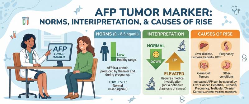
AFP is a protein produced in large quantities by the fetus and almost entirely absent from the blood after birth. In a healthy adult, its concentration is negligible. This is precisely why any significant AFP elevation in a non-pregnant adult woman or in a man is a serious signal that warrants further investigation. Here is what this marker measures, which diseases cause it to rise, and how to interpret the result correctly.
What Is Alpha-Fetoprotein (AFP) and Why Is It Ordered
AFP (alpha-fetoprotein) is a glycoprotein that serves as the principal transport protein of the embryo and fetus — it carries fatty acids, hormones and bilirubin, performing in the fetus the same role that albumin performs in the adult. Synthesised by the yolk sac and fetal liver, AFP peaks at the end of the first trimester and then gradually falls. After birth, levels drop sharply and reach adult values by 1–2 years of age.
In a healthy adult's blood, AFP is present only in trace amounts. A pathological rise in adults means one of two things: either cells have begun synthesising a protein characteristic of the embryonic period (typical of malignant tumours), or significant liver damage with regenerative activation has occurred.
Clinical roles of AFP:
- Treatment monitoring of hepatocellular carcinoma (HCC) — the primary liver cancer. AFP dynamics after surgery, transarterial chemoembolisation or systemic therapy are a key criterion of treatment response.
- HCC screening in high-risk patients — liver cirrhosis, chronic viral hepatitis B and C. AFP combined with liver ultrasound every 6 months is the standard surveillance protocol for these patients.
- Diagnosis and monitoring of germ cell tumours — non-seminomatous testicular and ovarian germ cell tumours. AFP is the first-line tumour marker in this setting.
- Prenatal screening — maternal serum AFP and amniotic fluid AFP are used to detect neural tube defects and chromosomal abnormalities in the fetus.
Like CEA, CA-125 and CA 19-9, AFP is part of the standard tumour marker panel and is ordered for specific clinical indications, not preventively.
AFP Normal Range
| Group | Normal range |
|---|---|
| Adult men | < 7.0 IU/mL (ng/mL) |
| Non-pregnant women | < 7.0 IU/mL (ng/mL) |
| Pregnant women | substantially higher — see trimester table below |
Different laboratories use different units and assay methods — always refer to the reference range on your own report.
AFP during pregnancy — normal ranges by gestational age:
| Gestational age | Normal (IU/mL) |
|---|---|
| 1–12 weeks | < 15 |
| 13–15 weeks | 15–60 |
| 16–19 weeks | 15–95 |
| 20–24 weeks | 27–125 |
| 25–27 weeks | 52–140 |
| 28–30 weeks | 67–150 |
| 31–32 weeks | up to 250 |
Interpretation outside pregnancy:
- < 7.0 IU/mL — normal
- 7–20 IU/mL — borderline; repeat measurement and clinical context assessment required
- 20–400 IU/mL — mild to moderate elevation. More often related to active liver injury than to tumour
- > 400 IU/mL — high; probability of a malignant process rises substantially
- > 1000 IU/mL in a patient with cirrhosis or hepatitis — diagnostic threshold for HCC under many international criteria
AFP as Tumour Marker: Hepatocellular Carcinoma and Germ Cell Tumours
Hepatocellular carcinoma (HCC) — the primary clinical application of AFP in adults. The marker is elevated in 60–70% of HCC patients. The key diagnostic threshold: AFP > 400 IU/mL in a patient with cirrhosis and a typical lesion on CT/MRI meets AASLD criteria for HCC diagnosis without biopsy. The higher the AFP, the worse the prognosis and the more advanced the tumour.
Critical limitation: 30–40% of HCCs are AFP-negative. A normal AFP does not exclude primary liver cancer. This is why HCC surveillance in cirrhosis is built on the combination of AFP and ultrasound, not AFP alone.
Non-seminomatous germ cell tumours of the testis — AFP is elevated in non-seminoma, embryonal carcinoma and yolk sac tumour. Pure seminoma does not raise AFP — if AFP is elevated in a patient diagnosed with pure seminoma, this indicates a non-seminomatous component and changes treatment. AFP is always measured alongside HCG: together they provide the complete marker profile for germ cell tumours.
Germ cell tumours of the ovary — yolk sac tumours and mixed germ cell tumours of the ovary raise AFP analogously to testicular tumours.
Hepatoblastoma — the malignant paediatric liver tumour. AFP is elevated in > 90% of patients, often reaching hundreds of thousands of IU/mL. AFP dynamics are the primary criterion of chemotherapy response in hepatoblastoma.
Liver metastases — AFP is mildly elevated in some patients with metastatic hepatic involvement of various origin, but it is not the marker of choice in this setting.
AFP in Cirrhosis, Hepatitis and Other Non-Cancer Causes
Mild AFP elevation in adults more commonly reflects benign liver disease than a tumour.
Liver cirrhosis — in active cirrhosis, AFP is chronically mildly elevated (usually < 200 IU/mL), a consequence of active hepatocyte regeneration: dividing cells resume synthesis of this embryonic protein. This is why a moderate AFP rise in a patient with cirrhosis cannot be unambiguously attributed to early HCC — serial measurement, ultrasound and CT are required.
Chronic viral hepatitis B and C — active inflammation and hepatocyte necrosis stimulate AFP synthesis. Levels are generally < 100 IU/mL; during flares, potentially higher. Successful antiviral therapy normalises AFP.
Acute hepatitis — massive hepatocyte necrosis (acute viral hepatitis, toxic liver injury) can produce a substantial AFP rise — sometimes reaching 200–400 IU/mL — as a regenerative response. This is a "false" elevation that resolves with recovery.
Pregnancy — physiologically elevated AFP is measured in maternal serum as part of prenatal screening.
Assessment of elevated AFP always includes liver function tests with ALT, AST, bilirubin and liver ultrasound.
How to Prepare for an AFP Blood Test
- Fasting — blood is drawn 8–12 hours after the last meal; required for reproducibility in serial monitoring.
- Active hepatitis or flares of chronic liver disease — during active inflammation, AFP will reflect regenerative activity rather than tumour status. Where possible, defer testing until remission.
- One laboratory for the entire monitoring course — different assay platforms produce non-comparable absolute values.
- High-dose biotin — discontinue 48 hours before the draw.
- A newly elevated AFP always requires simultaneous abdominal ultrasound: interpreting an isolated AFP rise without imaging is not possible.
AFP Interpretation: Trends in Cancer Monitoring
As with all tumour markers, the trend in AFP drives clinical decisions.
Normalisation after HCC treatment — after radical surgery or ablation, AFP should fall to normal within 4–8 weeks. Failure to normalise indicates residual tumour.
Rising AFP during cirrhosis surveillance — a doubling or more from baseline on two consecutive measurements 3–6 months apart requires contrast-enhanced CT to exclude HCC, even if the absolute value is not yet very high.
In germ cell tumours — AFP normalisation after chemotherapy is a criterion of complete response. Persistently elevated AFP after completing treatment is an indication for additional chemotherapy cycles or surgical removal of residual disease.
Rapidly rising AFP — a rise of tens of times over several weeks in a patient with established cirrhosis is a near-pathognomonic sign of malignant transformation.
Prenatal Screening: AFP in Pregnancy
In obstetrics, AFP is used as part of the combined prenatal screening — the "triple test" or "quadruple test" — alongside HCG, unconjugated oestriol and inhibin A.
Elevated maternal AFP (> 2.5 MoM — multiples of the median) indicates increased risk of neural tube defects (spina bifida, anencephaly), anterior abdominal wall defects (gastroschisis, omphalocele), multiple pregnancy, or fetal demise.
Low maternal AFP in combination with other markers is a sign of increased risk for Down syndrome (trisomy 21) and other chromosomal abnormalities.
Important: maternal serum AFP is a screening, not a diagnostic test. Any deviation from normal is an indication for detailed ultrasound and genetics consultation — it is not a diagnosis in itself.
When to See a Doctor
Urgent hepatology or oncology referral when:
- AFP > 400 IU/mL in a patient with cirrhosis or chronic hepatitis — emergency contrast-enhanced CT or MRI of the liver is required
- Any AFP elevation in a man with a testicular mass — requires immediate urological assessment
- Rising AFP during follow-up of HCC or germ cell tumour in remission
Scheduled gastroenterology or hepatology appointment when:
- AFP 20–400 IU/mL in a patient without oncological history — assess liver function and perform ultrasound
- Persistently mild AFP elevation in cirrhosis or chronic hepatitis — review HCC surveillance protocol
No emergency action needed:
- AFP mildly elevated during active acute hepatitis with a clear cause — retest after recovery
- AFP elevation in pregnancy — assessed by the obstetrician as part of prenatal screening
Elevated AFP is not a diagnosis — it is a signal for a structured diagnostic search. Do not interpret tumour markers independently; consult a physician.
This content is for informational purposes only and does not replace professional medical advice.
For informational purposes only
This article is for informational purposes only and does not constitute medical advice, diagnosis, or treatment. Please consult a healthcare professional for medical guidance.