Thyroid Cancer: Types, Symptoms, Diagnosis and Treatment
Reviewed by the LabReadAI medical team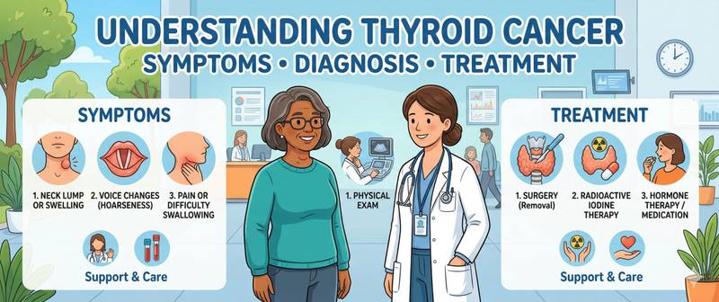
An ultrasound found a thyroid nodule. Or the doctor said "suspicious" and ordered a biopsy. Or a calcitonin test came back elevated. Thyroid cancer is one of the most heterogeneous groups of malignant diseases: some forms carry a 10-year survival above 95%, others are among the most aggressive tumours in oncology. Understanding the difference matters. This article covers the types of thyroid cancer, their symptoms, relevant blood tests, and treatment approaches.
Types of Thyroid Cancer and How They Differ
The thyroid gland contains two fundamentally different cell populations — follicular thyrocytes (which produce thyroxine and triiodothyronine) and parafollicular C-cells (which produce calcitonin). Three types of cancer arise from thyrocytes; one type from C-cells. This is not merely a classification exercise: the cell of origin determines the tumour's behaviour, its sensitivity to treatment, and the patient's prognosis.
Papillary thyroid cancer — the most common, comprising 80–85% of all cases. It grows slowly, metastasises predominantly to cervical lymph nodes, and rarely to lungs or bones. It readily absorbs radioactive iodine — making radioiodine therapy highly effective. Prognosis is excellent: the 10-year survival rate for localised disease exceeds 98%.
Follicular thyroid cancer — approximately 10–15%. Less likely to spread via lymphatics but prone to haematogenous dissemination: bone and lung metastases. Also sensitive to radioiodine. Prognosis is good when detected early; substantially worse with distant metastases.
Medullary thyroid cancer — about 5–10%. It arises from C-cells, does not absorb iodine, and does not respond to radioiodine therapy. Approximately 25% of cases are hereditary, linked to RET gene mutations. It frequently occurs as part of MEN-2A syndrome (combined with phaeochromocytoma and hyperparathyroidism) or MEN-2B. Prognosis is moderate: 10-year survival around 75%.
Anaplastic thyroid cancer — fewer than 2% of cases, but the most aggressive. Cells lose all differentiation; the tumour grows explosively. Median survival is 3–5 months. It occurs almost exclusively in patients over 60, frequently arising from a long-standing papillary or follicular cancer.
| Type | Proportion | Cell of Origin | Radioiodine | 10-Year Survival |
|---|---|---|---|---|
| Papillary | 80–85% | Thyrocyte | Yes | > 98% (localised) |
| Follicular | 10–15% | Thyrocyte | Yes | 85–95% |
| Medullary | 5–10% | C-cell | No | ~75% |
| Anaplastic | < 2% | Thyrocyte | No | < 10% |
Symptoms of Thyroid Cancer: When to Suspect It
The paradox of thyroid cancer is that most early forms produce no symptoms at all. Tumours are discovered incidentally: on routine neck ultrasound, during workup for another condition, or when the patient notices a lump while touching their throat.
A thyroid nodule is the most common initial finding. The critical point: the vast majority of thyroid nodules are benign. Population studies show nodules in 20–40% of adults on ultrasound, yet only 5–15% prove malignant. Nevertheless, every newly identified nodule requires assessment of cancer risk.
Features that raise suspicion for malignancy: rapid growth over weeks or months; a hard, "stony" consistency on palpation; fixation to surrounding tissues (does not move on swallowing); enlarged cervical lymph nodes; age below 20 or above 60; male sex (nodules are less frequent in men but more often malignant); family history of thyroid cancer or MEN syndrome.
Symptoms of local spread appear with large or invasive tumours: hoarseness (recurrent laryngeal nerve involvement), difficulty swallowing, breathing difficulty, neck pain radiating to the ear. These symptoms indicate locally advanced disease and require urgent oncological assessment.
Systemic symptoms are unusual in early thyroid cancer. In medullary cancer, excess calcitonin secretion occasionally causes diarrhoea — one of the rare paraneoplastic manifestations.
In hyperthyroidism associated with a functionally active cancer (rare, seen in some follicular cancers), tachycardia, weight loss, and sweating may be the presenting features.
Diagnosing Thyroid Cancer: Tests and Methods
Diagnosis combines imaging and laboratory testing. Neither alone provides a complete answer.
Thyroid ultrasound is the first and essential investigation for any suspected thyroid malignancy. It evaluates nodule characteristics using the TIRADS system (Thyroid Imaging Reporting and Data System): echogenicity, shape, margins, presence of microcalcifications, and vascularity. High-risk nodules (TIRADS 4–5) are referred for biopsy regardless of size.
Fine-needle aspiration biopsy (FNAB) — the primary method for obtaining tissue. Performed under ultrasound guidance, cells are aspirated with a fine needle for cytological examination. The Bethesda System classification (categories I–VI) determines subsequent management: from observation to immediate surgery.
Laboratory testing. TSH is the primary hormonal test. Its level helps assess thyroid function and provides indirect information about nodule type: functionally active ("hot") nodules are rarely malignant. A complete hormonal assessment uses the thyroid panel — TSH, free T4, and anti-TPO antibodies when indicated.
Tumour markers in thyroid cancer are used selectively. Thyroglobulin (Tg) is a marker for papillary and follicular cancer. It is poorly informative diagnostically (elevated in many benign conditions) but indispensable for post-operative surveillance: after total thyroidectomy, Tg should be undetectable. Any rise in Tg post-operatively is a signal of recurrence or metastasis.
Molecular-genetic testing of biopsy material is used in diagnostically indeterminate cytological categories (Bethesda III–IV): analysis of BRAF V600E, RET/PTC, RAS, and other mutations helps clarify malignancy before surgery.
Calcitonin and CEA: Markers for Medullary Thyroid Cancer
Medullary cancer is the only type of thyroid malignancy with highly specific biochemical blood markers. This makes it simultaneously one of the best-diagnosable and best-monitorable forms of the disease.
Calcitonin is the primary marker. C-cells — the origin of medullary cancer — retain the ability to secrete calcitonin in large quantities. A calcitonin level above 100 pg/mL in an adult is highly suspicious; above 500 pg/mL, the probability of medullary cancer approaches 100%. This is why European guidelines recommend measuring calcitonin in all patients with thyroid nodules before biopsy or surgical decision-making.
CEA (carcinoembryonic antigen) is the second marker for medullary cancer. It is less specific (rises in many cancers) but within the context of medullary disease its role is significant. The relative dynamics of calcitonin and CEA carry prognostic weight: when CEA rises faster than calcitonin, this indicates tumour dedifferentiation — a sign of more aggressive behaviour.
Post-operative monitoring in medullary cancer: after total thyroidectomy, both calcitonin and CEA should fall to undetectable levels. Persistently detectable calcitonin indicates incomplete tumour removal or occult metastases. Rising values on serial measurements over months are an indication for imaging to search for recurrence (neck and chest CT, MRI, scintigraphy).
For carriers of hereditary RET gene mutations (familial medullary cancer, MEN-2 syndromes), prophylactic thyroidectomy is recommended before cancer develops — in childhood or early adulthood depending on the specific mutation. This is the only type of thyroid cancer in which genetic testing of relatives is mandatory.
Treatment of Thyroid Cancer
Surgery is the cornerstone of treatment for all types of thyroid cancer. The extent of the operation depends on tumour type, size, and the presence of metastases.
Thyroidectomy (removal of the entire gland) is the standard for medullary cancer, anaplastic cancer, and most intermediate- and high-risk papillary and follicular cancers. For low-risk papillary cancer (tumour under 1 cm, no evidence of spread), hemithyroidectomy — removal of one lobe — is sometimes sufficient.
Radioiodine therapy (I-131) is used after total thyroidectomy for papillary and follicular cancer. Residual thyroid cells and metastases absorb the radioactive iodine and are destroyed from within. Efficacy is high: in most patients it eliminates microscopic metastases completely.
TSH-suppressive thyroxine therapy. After thyroidectomy, the patient takes L-thyroxine lifelong — not only as replacement therapy for hypothyroidism but also to suppress TSH to subnormal levels. TSH drives thyrocyte proliferation (including in tumour cells), so its suppression reduces recurrence risk in differentiated cancer.
Targeted therapy. For metastatic medullary cancer — tyrosine kinase inhibitors (vandetanib, cabozantinib) targeting RET and other molecular drivers. For progressive refractory papillary cancer — lenvatinib, sorafenib. For anaplastic cancer harbouring the BRAF V600E mutation — the combination of dabrafenib and trametinib.
External beam radiotherapy is used in medullary and anaplastic cancer, and for local recurrence in differentiated cancer patients.
Prognosis and Life After Thyroid Surgery
Most patients with papillary and follicular thyroid cancer live a normal lifespan. The five-year survival rate for localised papillary cancer approaches 100% — a figure better than virtually any other malignancy.
After total thyroidectomy, lifelong replacement therapy is required: with the gland removed, hypothyroidism is inevitable. Dose titration of L-thyroxine takes a few weeks, after which quality of life is generally indistinguishable from normal.
A possible surgical complication is hypoparathyroidism: during thyroid removal, the blood supply to the parathyroid glands may be compromised. This causes low blood calcium — numbness, muscle cramps, and spasms in the hands and feet. It is managed with calcium supplements and vitamin D.
Long-term surveillance for differentiated cancer includes: thyroglobulin and anti-Tg antibodies every 6–12 months; neck ultrasound annually for the first 3–5 years; I-131 scintigraphy if thyroglobulin rises. Recurrence risk is greatest in the first five years; after a 10-year disease-free period it is minimal.
When to See a Doctor Urgently
Seek prompt medical attention if:
- Calcitonin measured during a thyroid nodule workup exceeds 100 pg/mL — the threshold for high oncological concern; urgent endocrinology-oncology consultation is required.
- A neck mass is growing visibly over weeks: rapid growth is one of the primary red flags for malignancy.
- Hoarseness appears without a preceding cold or vocal strain — a possible sign of recurrent laryngeal nerve involvement by a growing tumour.
- Progressive difficulty swallowing or breathing develops alongside neck enlargement.
- Family history: a first-degree relative with medullary thyroid cancer or MEN-2 syndrome — an indication for prophylactic genetic testing and calcitonin measurement even without any symptoms.
- After thyroid surgery, thyroglobulin or calcitonin begins rising on serial measurements — a possible sign of recurrence.
This article is for informational purposes only and does not replace medical consultation. Result interpretation and diagnosis are made by a qualified physician.
For informational purposes only
This article is for informational purposes only and does not constitute medical advice, diagnosis, or treatment. Please consult a healthcare professional for medical guidance.