Lymphocytes in Blood: Normal Levels, Causes and Significance
Reviewed by the LabReadAI medical team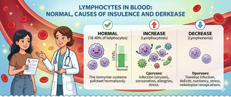
Lymphocytes are the intelligence center of the immune system. If neutrophils represent the rapid response to invasion, lymphocytes provide long-term memory and targeted elimination of specific pathogens. They recognize viruses and tumor cells, produce antibodies, and build immunological memory — the reason we don't suffer the same illness twice. Their deviation in a complete blood count is one of the most informative signals about the body's immune status.
What Lymphocytes Are and How They Are Classified
Lymphocytes are agranular mononuclear leukocytes — the second most numerous white blood cell type after neutrophils. They originate in the bone marrow but mature and specialize in different organs. By origin and function, lymphocytes fall into three main classes.
T-lymphocytes (thymus-derived) — mature in the thymus and constitute 60–80% of blood lymphocytes. They carry out cellular immunity:
- T-helper cells (CD4+) — the "conductors" of the immune response: they coordinate other immune cells and secrete cytokines
- Cytotoxic T-lymphocytes / T-killers (CD8+) — directly destroy virus-infected and tumor cells
- Regulatory T cells (Treg) — suppress excessive immune responses, preventing autoimmune reactions
B-lymphocytes — mature in the bone marrow and account for 10–15% of blood lymphocytes. They are the foundation of humoral immunity: when activated, they differentiate into plasma cells that produce antibodies (IgG, IgM, IgA, and others). B-lymphocytes are responsible for lasting antibody-based immunological memory.
NK cells (natural killers) — make up 5–20% of lymphocytes. They require no prior sensitization to a specific antigen: they identify and kill virus-infected and tumor cells through innate recognition — the frontline of antiviral and antitumor defense.
A standard complete blood count does not distinguish T, B, and NK lymphocytes — that requires immunophenotyping by flow cytometry. In routine CBC, lymphocytes are reported as a single combined figure.
Normal Lymphocyte Levels by Age
The proportion of lymphocytes versus neutrophils in healthy blood changes with age — a critical nuance in pediatric differential interpretation.
| Age | Lymphocytes (%) | Lymphocytes abs. (×10⁹/L) |
|---|---|---|
| Newborns | 20–40 | 2.0–11.0 |
| 1–6 months | 42–72 | 3.0–13.5 |
| 6 months – 1 year | 48–72 | 3.5–12.5 |
| 1–4 years | 38–72 | 2.5–9.5 |
| 4–6 years | 33–55 | 2.0–8.5 |
| 6–12 years | 25–50 | 1.5–7.0 |
| 12–18 years | 22–45 | 1.2–5.2 |
| Adults | 19–37 | 1.0–4.8 |
| Elderly > 65 years | 15–35 | 0.8–4.0 |
Two "physiological crossovers" — essential knowledge in pediatric hematology:
- First crossover (days 4–7 of life): neutrophils = lymphocytes (≈ 40–60% each)
- Second crossover (age 4–6 years): neutrophils = lymphocytes again (≈ 40–60% each)
Before the second crossover, lymphocytes predominate over neutrophils in healthy children — this is entirely normal, not lymphocytosis. Applying adult reference ranges to a child under 6 is a fundamental interpretive error.
In older adults, lymphocytes physiologically decline — a component of immune aging (immunosenescence): the naïve T-lymphocyte pool shrinks and the capacity to form new immune memory diminishes.
How to Prepare for a Lymphocyte Count
Lymphocytes are automatically calculated as part of the CBC with differential — no separate test is required.
- Blood drawn from a vein, morning, fasting or 3–4 hours after a light meal
- Physical exercise modestly reduces the relative lymphocyte percentage (due to neutrophil mobilization): avoid intense training for 24 hours beforehand
- Corticosteroids lower lymphocytes — always inform the physician when these are being taken
- Acute emotional stress transiently reduces lymphocytes via adrenaline-driven redistribution — repeat under calm conditions if the result is unexpected
- When chronic lymphoid pathology is suspected — refer for immunophenotyping (flow cytometry) to characterize T/B/NK subpopulations
Critical principle: the relative lymphocyte percentage must always be interpreted alongside the absolute count. 42% lymphocytes with total leukocytes of 3 × 10⁹/L is an absolute lymphopenia (1.26 × 10⁹/L), despite the apparently "high" percentage.
Causes of High Lymphocytes (Lymphocytosis)
Lymphocytosis — absolute count > 4.8 × 10⁹/L in adults — is divided into reactive (response to an external stimulus) and clonal (proliferation of a single pathological clone).
| Cause | Degree | Characteristic features |
|---|---|---|
| Viral infections (URTI, influenza, CMV) | Moderate | Transient; atypical lymphocytes |
| Infectious mononucleosis (EBV) | Significant | Atypical lymphocytes > 10%; pharyngitis; lymphadenopathy |
| Whooping cough (pertussis) | Significant (up to 15–20 × 10⁹/L) | Absolute lymphocytosis, especially in children |
| Toxoplasmosis | Moderate | Lymphadenopathy; atypical lymphocytes |
| Chronic lymphocytic leukemia (CLL) | Very high (> 20–50 × 10⁹/L) | Clonal; elderly; no infection symptoms |
| Lymphoma (leukemic phase) | High | Lymphadenopathy; B-symptoms |
| Thyrotoxicosis | Mild | Relative lymphocytosis |
| Convalescence after infection | Moderate | Transient; asymptomatic |
Infectious mononucleosis (Epstein-Barr virus) is the classic cause of acute lymphocytosis with atypical lymphocytes. Diagnostic picture: lymphocytes > 50% with atypical forms > 10%, pharyngitis, generalized lymphadenopathy, splenomegaly. Confirmed serologically (EBV VCA antibodies) or by PCR.
Chronic lymphocytic leukemia is the most common leukemia in adults over 60. Classic presentation: stable absolute lymphocytosis > 5 × 10⁹/L in an older patient without infection signs, without rising neutrophils or CRP. Requires immunophenotyping to confirm clonal B-cell origin.
Causes of Low Lymphocytes (Lymphopenia)
Lymphopenia — absolute count < 1.0 × 10⁹/L in adults — indicates suppression of cellular immunity.
| Cause | Mechanism | Characteristic features |
|---|---|---|
| Glucocorticoids | Redistribution to tissues and apoptosis | Dose-dependent; reversible |
| HIV/AIDS | Destruction of CD4+ T-lymphocytes | CD4 < 200 — severe immunodeficiency |
| Acute viral infections (early phase) | Redistribution to tissues | Transient; first 1–3 days |
| Severe bacterial infections / sepsis | Lymphocyte apoptosis during systemic inflammation | Parallel neutrophilia |
| Chemotherapy and radiotherapy | Direct destruction of proliferating cells | Proportional to treatment intensity |
| Autoimmune disease (SLE) | Anti-lymphocyte antibodies; consumption | Variable |
| Primary immunodeficiency | Impaired lymphocyte maturation | Recurrent infections from birth |
| Malnutrition and protein deficiency | Reduced production and function | In severe cachexia |
| Sarcoidosis | Lymphocyte sequestration in granulomas | Bilateral hilar lymphadenopathy |
HIV infection is the most clinically important cause of chronic progressive lymphopenia. The target is CD4+ T-helper cells: as HIV progresses their count falls. At CD4 < 200 cells/µL — AIDS stage, with risk of opportunistic infections (pneumocystis pneumonia, cytomegalovirus, cerebral toxoplasmosis). Unexplained absolute lymphopenia in a standard CBC in an adult is an indication for HIV testing.
Atypical Lymphocytes: What They Mean
Atypical (reactive, virocytes) lymphocytes are activated lymphocytes with altered morphology in response to antigenic stimulation — most often viral. In health they are absent or rare; a clinically significant finding is their presence at > 5–10% of all lymphocytes.
Morphological features: enlarged cell size, abundant basophilic cytoplasm, nucleus with fine chromatin structure. On Romanowsky-Giemsa staining — characteristic blue-grey cytoplasm with irregular margins.
Causes of atypical lymphocytes:
- Infectious mononucleosis (EBV) — the classic: atypical lymphocytes > 10–20%, also called "Downey cells"
- CMV infection — similar picture, but pharyngitis is less prominent
- Viral hepatitis — moderate numbers
- HIV (acute seroconversion phase) — together with lymphocytosis
- Allergic reactions and drug hypersensitivity
C-reactive protein during viral infections with atypical lymphocytes is often normal or only mildly elevated — in contrast to bacterial infections. This helps distinguish viral lymphocytosis from bacterial neutrophilia even before specific serological results return.
Atypical lymphocytes must be distinguished from blasts — malignant cells in leukemia. This is why significant lymphocytosis with atypical cells always warrants hematology consultation with expert morphological review of a stained blood smear.
When Lymphocyte Abnormalities Require Medical Attention
Scheduled visit to a doctor when:
- Lymphocytosis > 5 × 10⁹/L on repeat testing without signs of acute infection — rule out CLL and other lymphoproliferative conditions
- Lymphopenia < 1.0 × 10⁹/L not explained by corticosteroid use or acute infection
- Atypical lymphocytes > 10% with normal or moderately elevated total leukocytes
- Recurrent viral or fungal infections combined with chronic lymphopenia
Seek urgent care when:
- Lymphocytes > 30–50 × 10⁹/L — high probability of CLL or lymphoma leukemization
- Lymphopenia < 0.5 × 10⁹/L combined with fever — severe immunodeficiency with risk of opportunistic infection
- Lymphocytosis with atypical cells and signs of serious systemic disease: B-symptoms (night sweats, > 10% weight loss, fever > 38°C), marked lymphadenopathy, splenomegaly
This article is for informational purposes only and does not replace professional medical advice. Consult a GP or hematologist if your lymphocyte count is outside the normal range.
For informational purposes only
This article is for informational purposes only and does not constitute medical advice, diagnosis, or treatment. Please consult a healthcare professional for medical guidance.