Monocytes in Blood: Normal Range, High and Low Levels Explained
Reviewed by the LabReadAI medical team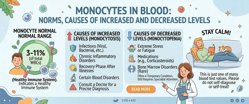
In a complete blood count, monocytes typically appear at the end of the white cell differential — and often pass unnoticed when neutrophils and lymphocytes are within range. Yet monocytes are unique cells with a double life: a few days in the bloodstream, then migration into tissues where they become macrophages — the immune system's principal long-term defenders and cleanup crews. Their elevation often signals a chronic threat rather than an acute one. Here's what the normal range looks like and what deviations mean.
What Are Monocytes and What Is Their Role
Monocytes are the largest cells among all leukocytes, reaching 14–21 µm in diameter — roughly twice the size of an average red blood cell. In health, they make up 2–10% of all white blood cells.
The bloodstream is only a transit stage for monocytes, lasting one to three days. They then migrate into tissues and differentiate — depending on the local environment — into macrophages (universal engulfers of pathogens and cellular debris) or dendritic cells (the primary antigen presenters for T lymphocytes). In macrophage form, they remain in tissues for months to years.
The functions of monocytes and their tissue derivatives span the entire immune cycle: phagocytosis of bacteria, viruses, and fungi; clearance of apoptotic cells and tissue debris; secretion of cytokines and chemokines that coordinate inflammation; participation in wound healing and tissue remodelling. Think of them as universal military engineers — they fight, build, and clean up after the battle.
Unlike neutrophils, which respond immediately and briefly, monocytes are cells of the sustained response. Their elevation more often signals a chronic process than an acute infection.
Normal Monocyte Counts by Age
Like other leukocytes, monocytes have age-specific reference ranges. In young children, their proportion is slightly higher than in adults.
| Age | Absolute Count (×10⁹/L) | Relative (%) |
|---|---|---|
| Newborns | 0.05–1.9 | 3–12 |
| Under 2 years | 0.05–1.1 | 3–10 |
| 2–12 years | 0.05–0.8 | 3–10 |
| Over 12 years | 0.04–0.8 | 2–10 |
| Adults | 0.04–0.8 | 2–10 |
Reference ranges may vary slightly between laboratories. Always use the values on your specific report.
Why Are Monocytes High? Monocytosis
Monocytosis — an absolute monocyte count above 0.8 × 10⁹/L — is not the most common finding in clinical practice, but it is diagnostically meaningful: it often points to conditions that other markers have not yet reflected.
Chronic infections are the classic territory of monocytosis. Tuberculosis is among the most prominent: in chronic TB, monocytes are persistently elevated because macrophages are the primary cells in which mycobacteria persist. The same applies to brucellosis, infective endocarditis, listeriosis, syphilis, and leishmaniasis. The defining feature of infectious monocytosis is duration — it persists for weeks and months until the primary focus is eliminated.
Recovery phase after acute infections — moderate monocytosis during convalescence is physiological. After neutrophils handle the acute phase, monocytes clear the inflammatory debris. This is transient and self-resolving.
Autoimmune diseases — systemic lupus erythematosus, rheumatoid arthritis, Crohn's disease, ulcerative colitis. In chronic autoimmune inflammation, monocytes and macrophages play a central role in sustaining the inflammatory cascade — and their blood levels are frequently elevated during flares.
Haematological malignancies — chronic myelomonocytic leukaemia (CMML) is defined by a persistent monocyte count above 1 × 10⁹/L as a diagnostic criterion. Some other haematological conditions also cause moderate monocytosis.
Sarcoidosis — a granulomatous disease in which granuloma formation depends directly on monocyte and macrophage activity.
Mild physiological monocytosis can occur post-surgery, with intense physical training, and in some apparently healthy individuals — without clinical significance.
Why Are Monocytes Low? Monocytopenia
Monocytopenia — an absolute count below 0.04 × 10⁹/L — is far less common than monocytosis and in most cases is a consequence of another condition or its treatment.
Corticosteroids are the most frequent cause. Glucocorticoids redistribute monocytes from blood into tissues and suppress their release from the bone marrow. This is a predictable and reversible treatment effect.
Aplastic anaemia — when all bone marrow cell lines are suppressed, monocyte production also falls. Monocytopenia in this setting is accompanied by reduced haemoglobin, neutropenia, and thrombocytopenia.
Hairy cell leukaemia — a rare but diagnostically important condition. The classic triad of pancytopenia, splenomegaly, and monocytopenia (often complete absence of circulating monocytes) is one of this disease's characteristic features.
Acute severe stress — transient monocytopenia occurs at the onset of some bacterial infections and during acute physiological stress. This is brief and clinically unimportant.
Reading Monocytes in the Context of the Full Differential
Monocytes are rarely interpreted in isolation — their meaning emerges in combination with other leukocytes and the clinical picture.
Several diagnostically significant patterns:
Monocytosis + normal neutrophils + normal lymphocytes — consider chronic infection (TB, brucellosis), sarcoidosis, or an autoimmune process.
Monocytosis + lymphocytosis + normal neutrophils — recovery phase after viral infection, mononucleosis in follow-up.
Persistent monocytosis > 1 × 10⁹/L on repeat tests without infection — grounds for haematology referral and CMML evaluation.
Monocytopenia + neutropenia + anaemia — pancytopenia requiring exclusion of aplastic anaemia and hairy cell leukaemia.
A complete assessment reads monocytes alongside neutrophils, lymphocytes, and ESR within a complete blood count with full differential.
How to Prepare for the Test
Monocytes are measured within a complete blood count with differential — no separate "monocyte test" exists. Preparation is standard.
Blood is drawn fasting after eight hours without food. Intense physical activity is best avoided for twelve hours beforehand — it has a moderate effect on monocyte counts. Tell your doctor about corticosteroid use: these drugs predictably suppress monocytes and may obscure the real picture.
If the purpose is monitoring a chronic condition (autoimmune or infectious), draw the test at the same time of day and under the same circumstances each time for comparable results.
When to See a Doctor
Schedule a routine appointment with a GP or haematologist if:
- Monocytosis above 1 × 10⁹/L is confirmed on repeat tests without active infection or a known autoimmune condition.
- Monocytopenia is combined with reduced counts in other cell lines — suggesting pancytopenia.
- Monocytosis persists more than four to six weeks after recovery from an acute illness.
- Monocytosis is discovered for the first time in an older adult without an obvious cause — CMML should be excluded.
Conclusion
Monocytes are cells of the long-term immune response. Moderate elevation during recovery from an infection is normal and self-resolving. Persistent monocytosis without acute inflammation signals a chronic process — infectious, autoimmune, or, less commonly, haematological. Monocytopenia combined with other low blood cell counts calls for urgent exclusion of serious bone marrow disease. In both cases, the key is a complete differential in serial testing — not a single isolated value.
This article is for informational purposes only. Interpreting test results and prescribing treatment is exclusively the responsibility of a physician.
For informational purposes only
This article is for informational purposes only and does not constitute medical advice, diagnosis, or treatment. Please consult a healthcare professional for medical guidance.