Infectious Mononucleosis: Symptoms, Diagnosis and Treatment
Reviewed by the LabReadAI medical team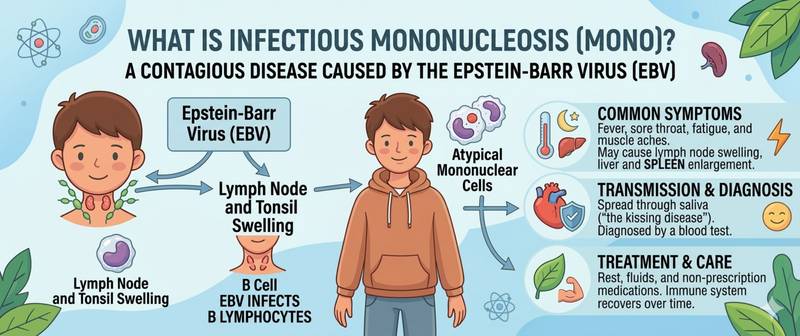
Severe sore throat, high fever, swollen lymph nodes — and the doctor says it is not ordinary tonsillitis but infectious mononucleosis. The diagnosis sounds unfamiliar, but the disease is far more common than most people realise: Epstein-Barr virus (EBV) infects around 95% of people during their lifetime. Let's look at how to distinguish mononucleosis from strep throat, which tests confirm the diagnosis, and why physical activity is genuinely dangerous with this illness.
What Is Infectious Mononucleosis and What Causes It
Infectious mononucleosis is an acute viral infection caused by Epstein-Barr virus (EBV, HHV-4) in 90% of cases. In 5–7% the causative agent is cytomegalovirus (CMV); rarely — other herpesviruses, toxoplasma, or primary HIV infection.
EBV is transmitted primarily through saliva — hence the unofficial name "kissing disease." It also spreads through shared utensils, toothbrushes, and blood transfusion. Incubation period: 4–6 weeks in adults, 1–2 weeks in children.
EBV is tropic to B-lymphocytes: it enters them via the CD21 receptor and drives their proliferation. In response, T-lymphocytes activate and attack the infected B-cells — these activated T-cells are what we see in the blood test as "atypical lymphocytes." They are not pathological cells in the classical sense; they are cytotoxic T-lymphocytes doing exactly their job.
After the acute infection, EBV persists lifelong in B-lymphocytes in a latent state. In people with a normal immune system, this causes no problems.
Symptoms of Infectious Mononucleosis
The classic presentation is a tetrad of symptoms found in most patients:
1. Fever — typically 38.5–40°C, lasting 1–2 weeks. May be continuous or wave-like.
2. Tonsillitis — severe sore throat. Tonsils are enlarged and covered with white or greyish-yellow exudate. Appears similar to streptococcal tonsillitis but does not respond to penicillins. In some patients the exudate is so extensive it nearly obstructs the pharynx.
3. Lymphadenopathy — enlarged lymph nodes, primarily cervical (especially posterior cervical — behind the sternocleidomastoid muscle). Axillary, inguinal, and mesenteric nodes may also enlarge. Nodes are tender, firm, and not fixed to the skin.
4. Splenomegaly — splenic enlargement, detected in 50–80% of patients. This finding defines the main restriction during illness. Rare but dangerous: splenic rupture with physical exertion.
Additional features:
- Hepatomegaly — in 10–15%; abnormal liver function tests — in most patients
- Rash — appears in 5–10% of patients without provocation
- Periorbital oedema (puffiness around the eyes) — a characteristic sign that distinguishes mononucleosis from other infections
- Palatal enanthem (petechiae on the soft palate)
- Profound fatigue — may persist for weeks to months after the acute phase
Mononucleosis and Antibiotics: A Critical Mistake to Know
Everyone should be aware: giving amoxicillin or ampicillin during mononucleosis causes a rash in 80–90% of patients. The mechanism is not a true allergic reaction — it is a specific immune phenomenon linked to EBV infection. Nevertheless, this rash is frequently and incorrectly documented as "amoxicillin allergy," permanently closing access to important antibiotics.
Why this happens: a doctor sees tonsillitis and prescribes penicillin thinking of streptococcus. In mononucleosis, tonsillitis is indeed accompanied by streptococcal co-infection in 15–30% of cases — but treating streptococcus during mononucleosis requires not amoxicillin, but other antibiotics — cephalosporins or macrolides if a penicillin-class drug is needed.
Diagnosing Mononucleosis from Blood Tests
Complete Blood Count with Differential
The complete blood count in mononucleosis produces a characteristic picture that is hard to mistake for another condition.
Leukocytosis — usually 10–20×10⁹/L, sometimes up to 30×10⁹/L. This may suggest bacterial infection — but the differential clarifies everything.
Lymphocytosis — lymphocytes make up 60–80% of all leukocytes (normal 20–40%). This is the most characteristic shift.
Atypical lymphocytes — present at more than 10% of all leukocytes. These are activated cytotoxic T-lymphocytes (CD8+) responding to EBV-infected B-cells. Under the microscope: large cells with wide basophilic cytoplasm and an "indented" nucleus. This finding is virtually pathognomonic for mononucleosis in the right clinical setting.
Mild thrombocytopenia — platelets fall to 100–150×10⁹/L in some patients. This resolves spontaneously without treatment.
ESR and CRP — moderately elevated, confirming active inflammation.
Biochemical Markers
Liver function tests are abnormal in most patients: ALT and AST elevated 2–10 times. This is EBV hepatitis — usually mild and self-limiting, but requires monitoring. Jaundice is rare (5%).
Serological Tests
Heterophile antibodies (Paul-Bunnell test, "monospot test") — rapid screening: specific IgM antibodies appearing in EBV infection. Sensitivity 85–90% in adults. Limitations: false-negative in the first week of illness and in children under 4 (often negative in this age group regardless of EBV).
Specific EBV serology (VCA IgM, VCA IgG, EA IgG, EBNA IgG) — the gold standard. Precisely identifies the phase of infection: acute, past, or reactivation.
| Marker | Acute infection | Past infection |
|---|---|---|
| VCA IgM | + | − |
| VCA IgG | + | + |
| EA IgG | + | − (usually) |
| EBNA IgG | − | + |
EBNA IgG appears 3–6 months after acute infection and persists for life — its presence confirms past infection.
The Main Danger: Splenic Rupture
Splenomegaly in mononucleosis is not just an incidental ultrasound finding. The enlarged, inflamed spleen becomes fragile and vulnerable to mechanical trauma. Splenic rupture occurs in approximately 0.1–0.5% of patients — but this is a life-threatening haemorrhage requiring emergency surgery.
Half of ruptures occur spontaneously, without obvious trauma. The rest follow physical contact, heavy lifting, coughing, or vomiting.
This is why the following are absolutely prohibited in mononucleosis:
- Contact sports (football, wrestling, basketball)
- Weightlifting and heavy lifting
- Running and any intense physical exertion
Activity restriction: minimum 3–4 weeks from illness onset; if splenomegaly is confirmed on ultrasound — until splenic size normalises.
Signs of splenic rupture: sudden severe pain in the left upper abdomen radiating to the left shoulder (Kehr's sign), rapidly worsening weakness, pallor, falling blood pressure — call emergency services immediately.
Treatment of Infectious Mononucleosis
There is no specific antiviral treatment proven effective in uncomplicated mononucleosis. Aciclovir and valaciclovir suppress EBV replication in the lab but have shown no clinical benefit in routine mononucleosis.
Treatment is symptomatic:
- Antipyretics and analgesics: paracetamol, ibuprofen. Aspirin is contraindicated in children and adolescents (risk of Reye's syndrome)
- Adequate hydration
- Rest and activity restriction
- Throat gargles with antiseptic solutions
- For severe tonsillar swelling with airway compromise risk — a short course of systemic corticosteroids
Antibiotics — only for confirmed concurrent bacterial infection (streptococcus confirmed by throat swab or rapid antigen test). Choice: cephalosporins or macrolides — never amoxicillin/ampicillin.
When to Seek Urgent Medical Attention
Call emergency services immediately for: sudden severe left upper abdominal pain — possible splenic rupture; worsening dyspnoea and stridor — airway obstruction from massively enlarged tonsils; high fever without improvement after 5 days in an infant.
See a doctor within 24 hours for: significant jaundice; neurological symptoms (severe headache, neck stiffness); rash after amoxicillin; temperature above 40°C not responding to antipyretics.
Summary
Infectious mononucleosis is a self-limiting viral illness: most patients recover within 2–4 weeks. The main dangers are splenic rupture during physical activity and the erroneous prescription of amoxicillin. The diagnostic key is a complete blood count with differential: lymphocytosis with atypical lymphocytes exceeding 10%, combined with the clinical picture, is nearly unmistakable. Post-mononucleosis fatigue may persist for months — this is normal and does not require specific treatment.
This article is for informational purposes only. Interpretation of test results and treatment decisions are the responsibility of a physician.
For informational purposes only
This article is for informational purposes only and does not constitute medical advice, diagnosis, or treatment. Please consult a healthcare professional for medical guidance.