How to Read a Blood Test: All CBC Indicators Explained
Reviewed by the LabReadAI medical team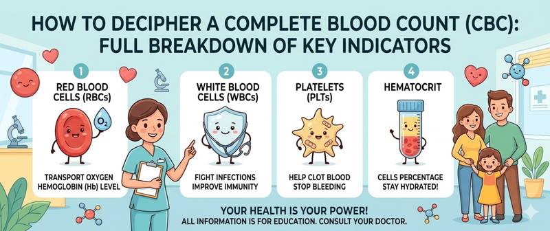
A results sheet with two dozen rows of numbers and upward or downward arrows — familiar territory for most patients. The complete blood count (CBC) is the most frequently ordered laboratory test: for any suspected infection, before surgery, at a routine check-up. But understanding what it actually says without an explanation is difficult. This article walks through every indicator of the complete blood count in order: what is measured, what counts as normal, and what deviations mean. The goal is not to replace a doctor — but to help you arrive at the appointment prepared.
How a Complete Blood Count Is Structured
A CBC consists of three interconnected components:
- Red cell indices: hemoglobin, red blood cells, haematocrit, MCV, MCH, MCHC, RDW — assess oxygen-carrying function and red cell morphology
- White cell indices: total leukocyte count and the differential (five-part count) — assess immune status
- Platelet indices: platelets, MPV — assess primary haemostasis
Results are expressed in absolute numbers (×10⁹/L or ×10¹²/L) or percentages. The laboratory prints a reference range next to every parameter — compare against this, not against internet averages: norms vary by equipment and reagent.
Read the panel as a whole: one isolated deviation is less significant than several indicators shifting in the same direction. Each indicator is best explained in context — not in isolation.
Hemoglobin and Red Blood Cells: Oxygen Transport
Hemoglobin (HGB, Hb) — the oxygen-carrying protein in red cells. The primary measure of blood oxygen transport. Normal: men 130–174 g/L, women 117–155 g/L, pregnant women from 105 g/L.
Low hemoglobin — anaemia. Graded by severity: mild (90 g/L to lower normal limit), moderate (70–89 g/L), severe (< 70 g/L). Symptoms: fatigue, breathlessness, pallor. Full breakdown of causes and treatment: low hemoglobin.
High hemoglobin — haemoconcentration (dehydration, polycythaemia). Thrombotic risk with chronic elevation.
Red blood cells (RBC) — normal: men 4.3–5.7 ×10¹²/L, women 3.8–5.1 ×10¹²/L. Reduction parallel to hemoglobin — anaemia. Isolated low RBC with normal hemoglobin — rare (minor thalassaemia).
Haematocrit (HCT) — the proportion of red cells in total blood volume, %. Normal: men 40–52%, women 35–47%. Low — anaemia or hypervolaemia; high — dehydration, polycythaemia.
MCV, MCH and RDW: Red Cell Morphology
These three parameters are the key to identifying the type of anaemia and narrowing the diagnostic workup immediately.
MCV (mean corpuscular volume) — normal 80–100 fL. The most important index:
- MCV < 80 fL (microcytosis) — iron deficiency anaemia, thalassaemia
- MCV 80–100 fL (normocytosis) — anaemia of chronic disease, haemolysis, acute blood loss
- MCV > 100 fL (macrocytosis) — vitamin B12 or folate deficiency, alcoholism, hypothyroidism
MCH (mean corpuscular haemoglobin) — normal 27–34 pg. Reduced in iron deficiency (hypochromia); elevated in macrocytic anaemias.
RDW (red cell distribution width / anisocytosis) — normal 11.5–14.5%. Elevated means red cells vary significantly in size. Low MCV + high RDW — classic iron deficiency anaemia (unlike thalassaemia, where RDW is normal).
Leukocytes and the Differential Count
Leukocytes (WBC) — white blood cells, the immune army. Normal in adults 4.0–9.0 ×10⁹/L. Elevated (leukocytosis) — infection, inflammation, stress, blood malignancy. Reduced (leukopenia) — viral infections, immunodeficiency, bone marrow disease.
The total leukocyte count without a differential is only the starting point — this indicator alone cannot be explained without breaking it down further. A doctor always evaluates the five-part differential — the leukocyte formula.
Neutrophils — normal 48–78% (1.8–7.7 ×10⁹/L). The most numerous fraction. Elevated (neutrophilia) — bacterial infection, inflammation, stress, corticosteroids. A "left shift" — appearance of immature forms (band cells > 5%) — signals severe infection or sepsis. Reduced (neutropenia) — viral infections, cytotoxics, aplastic anaemia.
Lymphocytes — normal 19–37% (1.0–3.5 ×10⁹/L). Elevated (lymphocytosis) — viral infections, chronic lymphocytic leukaemia. Reduced (lymphopenia) — immunodeficiency, HIV, prolonged steroid therapy.
Monocytes — normal 3–11% (0.09–0.6 ×10⁹/L). Elevated — chronic infections (tuberculosis, brucellosis), inflammatory bowel disease, recovery phase.
Eosinophils — normal 1–5% (0.02–0.44 ×10⁹/L). Elevated — allergic disease, parasitic infections, drug reactions.
Basophils — normal 0–1% (0–0.08 ×10⁹/L). Elevated in allergy, hypothyroidism, chronic myelogenous leukaemia.
Platelets and MPV: Haemostasis
Platelets (PLT) — blood cells that form the primary haemostatic plug. Normal 150–400 ×10⁹/L. Reduced (thrombocytopenia < 150) — bleeding risk. Below 50 — spontaneous haemorrhage; below 20 — life-threatening. Causes: immune thrombocytopenia, DIC, leukaemia, cirrhosis. Elevated (thrombocytosis > 400) — inflammation, iron deficiency, post-splenectomy, myeloproliferative disorders.
MPV (mean platelet volume) — normal 7–12 fL. Large platelets (high MPV) — young, active cells: seen after blood loss, in immune thrombocytopenia. Small platelets (low MPV) — aplastic anaemia, chemotherapy.
ESR: The Inflammation Marker
ESR (erythrocyte sedimentation rate) — an indirect marker of systemic inflammation. Normal: men up to 15 mm/h, women up to 20 mm/h (Westergren method). Higher limits in older adults.
ESR is non-specific: it rises with any inflammation, infection, autoimmune condition, malignancy — and even in normal pregnancy. On its own, an elevated ESR does not reveal what is inflamed — only that inflammation exists.
High ESR (> 50–60 mm/h) with otherwise normal CBC — a red flag: requires a search for the cause: malignancy, myeloma, systemic connective tissue disease. Mildly elevated ESR (20–40 mm/h) with good wellbeing is often a physiological variant or residual from a recent infection.
ESR responds slowly — it may remain elevated for weeks after recovery. It is not used to assess the acuity of a current condition — only for tracking trends.
How to Read Your Blood Count Report: A Step-by-Step Approach
- Use the laboratory's reference range, not internet averages.
- Distinguish isolated from combined abnormalities. A single mild deviation is less significant than several parameters shifting together.
- Assess the trend over time. Hemoglobin 118 g/L in a woman whose previous value was 135 g/L is significant, even if technically "within normal."
- Match results to symptoms. Leukocytosis with a fever of 39 °C is a very different context from the same leukocytosis after an intense gym session with no symptoms.
- One test is not a diagnosis. Any significant deviation warrants a repeat test and a medical consultation.
Common Patterns of Deviation and What They Mean
Several clinically important patterns that a doctor recognises at a glance — each explained by the combination of indicators rather than a single value:
Low hemoglobin + low MCV + high RDW → iron deficiency anaemia — the most common pattern in practice.
Low hemoglobin + high MCV + normal leukocytes and platelets → B12 or folate deficiency.
High leukocytes + neutrophilia + left shift → bacterial infection or sepsis.
High leukocytes + lymphocytosis + atypical lymphocytes → viral infection (infectious mononucleosis, CMV).
Low leukocytes + low platelets + low hemoglobin → pancytopenia — suppression of all bone marrow cell lines. Requires urgent haematology workup.
High ESR + normal leukocytes → chronic inflammation, autoimmune process, malignancy.
High platelets + low MCV + low hemoglobin → iron deficiency anaemia (reactive thrombocytosis in iron deficiency).
When CBC Abnormalities Require Urgent Medical Attention
Most moderate deviations warrant a scheduled appointment. But when critical indicators are present, the urgency is explained by the risk of immediate complications:
- Hemoglobin < 70 g/L regardless of symptoms
- Leukocytes > 30 × 10⁹/L or < 1.0 × 10⁹/L
- Platelets < 30 × 10⁹/L — haemorrhage risk
- Blast cells on the smear — sign of acute leukaemia
- Pancytopenia — simultaneous reduction of all three cell lines
- Neutrophils < 0.5 × 10⁹/L — agranulocytosis
- Any critical deviation in a pregnant woman
This content is for informational purposes only and does not replace professional medical advice.
For informational purposes only
This article is for informational purposes only and does not constitute medical advice, diagnosis, or treatment. Please consult a healthcare professional for medical guidance.