High White Blood Cell Count: Causes of Leukocytosis and What to Do
Reviewed by the LabReadAI medical team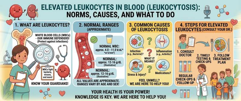
You received your complete blood count and found that your white blood cell count is above normal. The first thought is usually infection. In most cases that is correct: leukocytosis is the immune system's signal that it is fighting something. But sometimes elevated white blood cells reflect a medication, stress, pregnancy, or — in rare cases — a blood malignancy. What it means and how serious it is depends not on the total number alone, but on the full differential count together with the clinical picture. This article covers normal ranges, causes of leukocytosis by cell fraction, and when the elevation requires urgent attention.
Normal White Blood Cell Count: When Is a Value Considered High?
Leukocytes — white blood cells — are the immune system's primary weapons against infection, inflammation, and foreign cells. Leukocytosis is a total white blood cell count above the upper limit of normal.
Normal white blood cell count:
| Category | Normal (× 10⁹/L) |
|---|---|
| Adults | 4.0–9.0 |
| Pregnant women | 6.0–11.0 |
| Infants under 1 year | 6.0–17.5 |
| Children 1–6 years | 5.0–15.5 |
| Children 6–12 years | 4.5–13.5 |
| Adolescents | 4.5–11.0 |
Leukocytosis grades:
- Mild: 10–15 × 10⁹/L — often physiological or reactive
- Moderate: 15–30 × 10⁹/L — typically infectious or inflammatory
- Severe: above 30 × 10⁹/L — mandatory workup to rule out haematological pathology
- Hyperleukocytosis: above 100 × 10⁹/L — critical, characteristic of leukaemia
A key principle: the total white cell count without a differential is only the starting point. A doctor always evaluates the leukocyte differential to determine which cell population is elevated — this fundamentally changes the diagnostic picture.
Physiological Leukocytosis: When Elevation Is Normal
Not all leukocytosis signals disease. Several conditions cause physiological white cell elevation that resolves on its own and requires no treatment.
Alimentary leukocytosis — after a large meal, especially high in protein. This is why a blood count must be drawn strictly fasting: eating 2–3 hours before the draw can falsely elevate white cells by 20–30%.
Exercise-induced leukocytosis — after intense physical exertion, white cells redistribute from tissue reservoirs into the bloodstream. Levels return to normal within hours.
Emotional leukocytosis — stress, fear, and pain trigger catecholamine release, mobilising white cells from the bone marrow. Testing during acute emotional distress risks a false result.
Pregnancy — physiological leukocytosis from the second trimester onward. Normal in pregnancy is up to 11.0 × 10⁹/L; levels may reach 12–15 × 10⁹/L before delivery.
Smoking — chronic tobacco smoke exposure causes a persistent mild leukocytosis from chronic airway inflammation.
Newborns — white cell counts of 10–30 × 10⁹/L are normal in the first days of life, gradually declining toward school age.
Causes of Leukocytosis in Adults: From Infections to Malignancy
Pathological leukocytosis always has a cause. The pattern of elevation and the affected fraction are the key to finding it.
Infections — the most common cause:
- Bacterial infections (pneumonia, pyelonephritis, tonsillitis, sepsis) produce neutrophilia with a left shift — appearance of immature neutrophils (band cells, metamyelocytes). The more severe the infection, the more pronounced the shift.
- Viral infections cause lymphocytosis with normal or mildly elevated total white cells. A classic example: infectious mononucleosis, in which lymphocytes rise sharply with atypical forms appearing on the smear.
- Parasitic infestations and allergies — eosinophilia: leukocytosis driven specifically by the eosinophil fraction.
Inflammatory and autoimmune diseases:
- Rheumatoid arthritis, systemic lupus erythematosus, inflammatory bowel disease — moderate neutrophilia without infection.
- Acute myocardial infarction — pronounced neutrophilia in the first 12–24 hours, reflecting tissue necrosis.
Medications:
- Glucocorticoids (prednisone, dexamethasone) — the most common pharmacological cause of neutrophilia.
- Epinephrine and beta-agonists — mobilise the marginal neutrophil pool.
- Lithium — stimulates bone marrow granulopoiesis.
- G-CSF (granulocyte colony-stimulating factor) — used in oncology; produces pronounced neutrophilia as its intended effect.
Oncological and haematological causes — considered with persistent leukocytosis of no apparent cause:
- Acute leukaemia — chaotic blast-cell leukocytosis, anaemia, thrombocytopenia.
- Chronic myelogenous leukaemia — gradually rising leukocytosis to 50–300 × 10⁹/L, splenomegaly.
- Chronic lymphocytic leukaemia — persistent lymphocytosis in older adults.
- Myeloproliferative disorders — polycythemia vera, essential thrombocythaemia.
Stress and trauma:
- Major burns, severe trauma, post-operative state — neutrophilia as a marker of tissue damage.
- Splenic or intestinal infarction — acute pronounced neutrophilia.
Types of Leukocytosis by Elevated Fraction
Knowing which fraction is elevated immediately narrows the list of possible diagnoses.
Neutrophilia (neutrophils above 7.5 × 10⁹/L) — the most common type of leukocytosis. Indicates bacterial infection, inflammation, stress, or corticosteroid use. A left shift (band cells > 5%) signals active bacterial infection or sepsis.
Lymphocytosis (lymphocytes above 3.5 × 10⁹/L) — viral infections, chronic lymphocytic leukaemia, tuberculosis, toxoplasmosis. Atypical lymphocytes on the smear are characteristic of infectious mononucleosis and cytomegalovirus infection.
Monocytosis (monocytes above 0.8 × 10⁹/L) — chronic infections (tuberculosis, brucellosis), inflammatory bowel disease, recovery phase of acute infections.
Eosinophilia (eosinophils above 0.5 × 10⁹/L) — allergic diseases (bronchial asthma, atopic dermatitis), parasitic infections, drug reactions, malignancies.
Basophilia (basophils above 0.1 × 10⁹/L) — allergies, hypothyroidism, chronic myelogenous leukaemia.
High White Blood Cells in Children: Ranges and Interpretation
In children, normal white cell counts are substantially higher than in adults, and change with age. This means that an "elevated" value in a child must be compared against age-specific norms, not adult references.
Characteristics of childhood leukocytosis:
- In children under 5, lymphocytes physiologically predominate (up to 60%), not neutrophils — the physiological "crossover." Moderate lymphocytosis in a 2–4 year old is a normal variant, not a sign of disease.
- Reactive leukocytosis during a respiratory illness in children often reaches 15–20 × 10⁹/L — not alarming if symptoms are mild and the differential shows a viral pattern.
- In newborns, even 20–30 × 10⁹/L can be within the normal range.
- Persistent unexplained leukocytosis in a child without infectious symptoms warrants a paediatric haematology consultation.
For correct interpretation in children, see complete blood count in children with detailed age-specific reference ranges.
High White Blood Cells in Pregnancy
Leukocytosis during pregnancy is physiological, driven by increased immune activity, expanded blood volume, and cortisol release. The normal upper limit in pregnancy is 11.0 × 10⁹/L.
Nevertheless, leukocytosis in pregnancy requires attention in several situations:
- White cells above 15 × 10⁹/L — infection must be excluded: pyelonephritis, chorioamnionitis, appendicitis.
- Pronounced neutrophilia with a left shift — signs of active bacterial inflammation.
- Any rise in white cells combined with fever, pain, or urinary abnormalities — immediate obstetric consultation.
When High White Blood Cells Require Urgent Medical Attention
A mild transient leukocytosis after a respiratory illness or physical exertion does not require emergency action. But several situations call for immediate consultation:
- White cells above 30 × 10⁹/L without an identifiable infectious cause
- Hyperleukocytosis above 100 × 10⁹/L — medical emergency
- Leukocytosis combined with anaemia and thrombocytopenia — tri-lineage involvement
- Blast cells found on the blood smear — sign of acute leukaemia
- Persistently elevated white cells on two or more tests without infection symptoms
- Enlarged lymph nodes, spleen, or liver combined with leukocytosis
- Pronounced leukocytosis in a child without an obvious cause
- Neutrophilia with a left shift + fever above 38.5 °C + deteriorating condition — possible sepsis
For moderate leukocytosis with respiratory symptoms and good overall wellbeing — a repeat test 2–3 weeks after recovery. If white cells remain elevated, a scheduled GP visit is appropriate.
Conclusion
Elevated white blood cells are a symptom, not a disease. Behind leukocytosis can stand dozens of conditions ranging enormously in severity — from a common cold to haematological malignancy. The key to correct interpretation is looking beyond the total count to the differential, the trend, the clinical symptoms, and the patient's history. Moderate leukocytosis during an infection that normalises after recovery is the expected immune response. Persistent elevation without an explainable cause warrants a full diagnostic workup.
This content is for informational purposes only and does not replace professional medical advice.
For informational purposes only
This article is for informational purposes only and does not constitute medical advice, diagnosis, or treatment. Please consult a healthcare professional for medical guidance.