Vitamin A Blood Test: Normal Levels, Deficiency and Toxicity
Reviewed by the LabReadAI medical team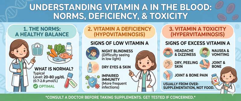
The first sign many patients notice themselves is difficulty adapting their eyes in dim light: a familiar room becomes hard to navigate, and oncoming headlights are blinding in a way they never used to be. Impaired twilight vision is not merely an inconvenience — it is an early biomarker of retinol deficiency. A blood test for vitamin A can reveal the problem before more serious consequences take hold.
What Vitamin A Is and Its Different Forms
Vitamin A is a group of fat-soluble compounds that share a common biological action. In the body it exists in two primary forms: retinol — preformed vitamin A obtained from animal foods — and carotenoids, the plant-derived provitamins A that are converted into retinol in the intestinal wall. The most familiar carotenoid is beta-carotene, responsible for the orange colour of carrots and pumpkin.
The biological functions of vitamin A span three critical systems. The first is vision: retinol is a structural component of rhodopsin, the light-sensitive pigment of retinal rod cells. Rhodopsin captures photons in dim light and converts the light signal into a nerve impulse. This is precisely why retinol deficiency impairs twilight and night vision before any other symptom appears.
The second system is immunity. Vitamin A is required for the differentiation and maturation of T-lymphocytes and mucosal epithelial cells, which form the primary barrier against infection. Retinol deficiency dramatically increases mortality from measles and diarrhoeal disease in children — this is among the most robust findings in nutritional epidemiology.
The third is cellular growth and differentiation. Through nuclear receptors (RAR and RXR), vitamin A regulates the expression of hundreds of genes governing cell division and specialisation in the skin, respiratory epithelium and gastrointestinal tract. In deficiency, normal columnar epithelium is replaced by keratinising squamous cells — a process called squamous metaplasia.
Retinol is transported in blood by retinol-binding protein (RBP), the synthesis of which requires both zinc and dietary protein. This is why zinc deficiency can lower plasma retinol even when hepatic stores are normal: the liver holds vitamin A but cannot release it without RBP.
Normal Vitamin A Blood Levels
Vitamin A in blood is measured as plasma retinol by high-performance liquid chromatography (HPLC).
| Status | Plasma retinol level |
|---|---|
| Normal | 1.05–3.50 µmol/L (30–100 µg/dL) |
| Suboptimal | 0.70–1.05 µmol/L |
| Deficient | < 0.70 µmol/L |
| Severely deficient | < 0.35 µmol/L |
| Potentially toxic | > 3.50 µmol/L |
Reference ranges vary between laboratories — always compare against the values printed on your own report.
An important interpretive caveat: plasma retinol is not the most sensitive marker of body stores. The liver holds 85–90% of total vitamin A and sustains normal plasma levels for a prolonged period by gradually drawing down hepatic reserves. A fall in plasma retinol occurs only when hepatic stores are already substantially depleted. More precise assessments — the relative dose response (RDR) test and isotope dilution — are used in research, not routine clinical practice.
Reference ranges are lower in children than in adults. In pregnancy, levels decline modestly in the third trimester as retinol is transferred to the fetus. Acute-phase proteins released during any inflammatory condition reduce hepatic RBP synthesis, falsely lowering plasma retinol. Vitamin A should never be interpreted during an active infection, pneumonia or other inflammatory illness.
How to Prepare for a Vitamin A Blood Test
Vitamin A is a fat-soluble analyte that is stable under correct storage conditions but sensitive to light exposure and dietary status before collection.
Preparation:
- Draw blood strictly fasting — 8–12 hours without food
- Stop vitamin A supplements and multivitamins 24–48 hours beforehand
- For one week before, avoid abnormally large amounts of carotenoid-rich foods (e.g. litres of carrot juice)
- No smoking for 2 hours before the draw
The blood tube must be shielded from light immediately after collection. Retinol degrades rapidly on exposure to ultraviolet light — the laboratory must handle the sample under protected conditions. For a comprehensive picture of nutritional status, a combined vitamin panel provides simultaneous assessment of multiple fat-soluble and water-soluble vitamins, eliminating the risk of an isolated false result and revealing co-existing deficiencies.
Causes and Symptoms of Vitamin A Deficiency
Vitamin A deficiency remains the leading preventable cause of childhood blindness globally: the WHO estimates 250 million pre-school children are affected. In high-income countries clinical deficiency is rare, but suboptimal levels are found in 5–10% of adults — particularly in at-risk groups.
Main causes:
- A diet low in retinol: strict veganism without dietary planning, monotonous diets in older adults
- Impaired fat absorption: coeliac disease, Crohn's disease, cystic fibrosis, chronic pancreatitis, bariatric surgery. Since vitamin A is fat-soluble, absorption collapses when fat digestion is impaired regardless of dietary intake
- Zinc deficiency: without adequate zinc, the liver cannot release stored retinol via RBP
- Liver cirrhosis: a damaged liver loses its capacity to store vitamin A and synthesise RBP
- Chronic alcoholism: impaired absorption and competition between alcohol and retinol for shared enzyme systems
- Prematurity: the fetus accumulates most of its vitamin A reserves during the final trimester
Clinical stages:
Early deficiency presents as impaired twilight vision — nyctalopia ("night blindness"). This is fully reversible with repletion. Simultaneously, resistance to infections declines: patients experience more frequent respiratory and gastrointestinal infections, with prolonged recovery.
Moderate deficiency adds skin and mucosal involvement: hyperkeratosis (dry, flaking skin, "goose flesh" on the upper arms and thighs), conjunctival dryness and reduced tear production. The skin loses elasticity and becomes prone to fissuring. Thinning of the respiratory and intestinal mucosa increases vulnerability to infection.
Severe deficiency leads to xerophthalmia — progressive corneal disease. Dryness advances to Bitot's spots (greyish plaques on the conjunctiva), then to keratomalacia — softening and perforation of the cornea with irreversible blindness. At this stage, ocular damage is permanent.
Hypervitaminosis A: The Risks of Excess
Vitamin A is one of the few vitamins that accumulates in the liver and can reach toxic concentrations. Unlike water-soluble vitamins excreted rapidly by the kidneys, retinol is stored, and chronic overdose leads to serious consequences.
Acute hypervitaminosis A develops after a single very large dose (> 200,000 IU in adults). It was historically documented in polar explorers who consumed polar bear or seal liver. Manifestations include severe headache, nausea, vomiting, vertigo and raised intracranial pressure.
Chronic hypervitaminosis A is far more common and more insidious. It develops with sustained supplementation exceeding 10,000 IU/day over months to years. Symptoms accumulate gradually: dry and peeling skin (especially the lips), hair loss, bone and joint pain, chronic fatigue, hepatomegaly and elevated liver transaminases.
The principal long-term risk is hepatotoxicity — fibrosis progressing to cirrhosis with very high doses sustained over years. The second serious risk is osteoporosis: chronically elevated retinol activates osteoclasts and suppresses osteoblasts, reducing bone mineral density and raising fracture risk. This effect is most pronounced in older women.
Teratogenicity is an absolute contraindication to high doses in the first trimester of pregnancy. Retinoic acid is a potent regulator of embryogenesis: supplemental doses above 10,000 IU/day during pregnancy are associated with malformations of the heart, central nervous system and face. Carotenoids from plant foods are safe: their conversion to retinol in the intestine is tightly regulated and does not cause hypervitaminosis A.
Carotenaemia — yellow-orange skin discolouration from dietary beta-carotene excess — is harmless and fully reversible. It is not hypervitaminosis A, as carotenoids are not converted to toxic retinol quantities.
Vitamin A and Other Fat-Soluble Vitamins
Vitamin A does not act in isolation. As a fat-soluble compound, it shares absorption and metabolic pathways with other fat-soluble vitamins — and between them exist both synergies and competition.
Vitamin D and vitamin A share the RXR nuclear receptor. When both are simultaneously deficient, each deficiency worsens the other; correcting one without the other produces an incomplete response. This is particularly relevant for older patients and those in northern latitudes where both vitamins are systemically low.
Vitamin E — the primary fat-soluble antioxidant — protects retinol stores in cell membranes from oxidative degradation. When vitamin E is deficient, retinol is consumed more rapidly. Vitamin K2 governs calcium distribution in bone; its deficiency in the presence of excess vitamin A amplifies bone mineral loss.
In practice: supplementing vitamin A in isolation without assessing the status of other fat-soluble vitamins is incomplete management. All fat-soluble vitamins also share a dependency on dietary fat for absorption — taking supplements on an empty stomach or with fat-free food dramatically reduces bioavailability.
When to See a Doctor
Testing for vitamin A is warranted when one or more of the following apply:
- impaired twilight vision or abnormal sensitivity to bright light;
- dry, flaking skin — particularly on the upper arms and thighs — without another explanation;
- frequent respiratory or gastrointestinal infections;
- diagnosed fat malabsorption: coeliac disease, Crohn's disease, chronic pancreatitis;
- long-term supplementation with vitamin A above 5000 IU/day;
- pregnancy planning or first trimester — to confirm a safe circulating level.
Self-prescribing high-dose retinol supplements without a blood test is hazardous: chronic hypervitaminosis develops gradually and its early symptoms are non-specific. For a comprehensive evaluation of nutritional status, ask your doctor for a combined vitamin panel — a single assessment that reveals deficiencies and excesses across multiple vitamins simultaneously.
This content is for informational purposes only and does not replace professional medical advice.
For informational purposes only
This article is for informational purposes only and does not constitute medical advice, diagnosis, or treatment. Please consult a healthcare professional for medical guidance.