Ankylosing Spondylitis: Symptoms, Diagnosis and Treatment
Reviewed by the LabReadAI medical team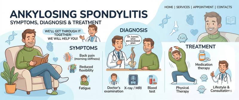
Low back pain in a young man that worsens at rest and eases with movement. Morning stiffness that lasts more than an hour. This is not "disc disease" — this is the classic presentation of ankylosing spondylitis (AS), a chronic inflammatory disease of the spine. In early disease, with timely diagnosis and treatment, most patients maintain normal mobility and quality of life; delayed treatment leads to irreversible spinal fusion.
What Is Ankylosing Spondylitis (Sacroiliitis) and How Does It Develop
Ankylosing spondylitis is part of the seronegative spondyloarthritis group — inflammatory diseases of the axial skeleton and joints that are not associated with rheumatoid factor. The primary targets are the sacroiliac joints (sacroiliitis) and the spine; peripheral joints, eyes, heart, and bowel are less commonly affected.
Inflammation in AS develops at entheses — the sites where tendons, ligaments, and joint capsules attach to bone. Chronic entheseal inflammation triggers pathological bone formation: fibrous and ultimately bony "bridges" — syndesmophytes — form between vertebrae and progressively fuse the spinal column. The end result of untreated progressive disease is the "bamboo spine": complete fusion of the entire vertebral column.
The key genetic marker is the HLA-B27 antigen: present in 90–95% of AS patients, while occurring in only 6–8% of the general population. However, HLA-B27 is not itself a diagnosis: only 1–5% of carriers ever develop AS. The disease occurs 2–3 times more often in men and typically begins between ages 15 and 35.
Symptoms of Ankylosing Spondylitis: Inflammatory Back Pain and Morning Stiffness
Inflammatory low back pain — the hallmark of AS and its primary distinction from mechanical back pain. Criteria for inflammatory pattern: onset before age 45; gradual onset; improvement with physical activity and movement; no relief with rest; nocturnal worsening (especially in the second half of the night). Morning stiffness lasting more than 30 minutes is an almost universal feature.
Mechanical pain from disc degeneration, by contrast, worsens with movement, improves with rest, and does not wake the patient at night.
Sacroiliitis — inflammation of the sacroiliac joints — is frequently the first manifestation, appearing years before spinal involvement. It presents as buttock pain, often alternating sides, radiating to the thighs.
Restricted spinal mobility develops as the disease progresses. The finger-to-floor distance on forward bending increases; chest expansion decreases (normal > 5 cm).
Peripheral arthritis occurs in 25–35% of patients: large lower limb joints — hip and knee — are most commonly affected. Hip involvement carries the greatest functional disability burden in AS.
Enthesitis — pain at tendon insertion sites: the Achilles tendon, plantar fascia, and costochondral junctions are the most typical locations.
Uveitis — acute inflammation of the uveal tract — develops in 30–40% of AS patients. It presents with sudden eye pain, redness, and photophobia. Immediate ophthalmological treatment is required to prevent vision loss.
Extra-articular manifestations: inflammatory bowel disease (Crohn's disease, ulcerative colitis) coexists with AS in 5–10% of patients; psoriasis in approximately 10%.
Diagnosing Ankylosing Spondylitis: Tests and Methods
The diagnosis of AS is clinical, based on a combination of symptoms, laboratory findings, and imaging. The Modified New York Criteria (1984) and the ASAS criteria for axial spondyloarthritis (2009) remain the standard frameworks.
HLA-B27 — a genetic marker. A positive result substantially raises the probability of AS in the appropriate clinical context, but is not a diagnostic criterion on its own. In a patient with inflammatory back pain and positive HLA-B27, the probability of AS is approximately 50%.
Inflammatory markers. C-reactive protein and ESR are elevated in 40–70% of patients with active AS — but in a subset of patients (particularly those with predominantly axial disease) these markers may remain normal even with high disease activity. Normal CRP and ESR do not exclude AS — clinical judgement outweighs laboratory findings.
Rheumatoid factor and anti-CCP antibodies are typically negative in AS — this is why AS is called a "seronegative" spondyloarthritis. These tests are ordered primarily to differentiate AS from rheumatoid arthritis.
Complete blood count — mild normochromic anaemia in active disease, thrombocytosis as an acute-phase response.
MRI of the sacroiliac joints — the most sensitive tool for early diagnosis. It detects active inflammation (bone marrow oedema) before structural changes are visible on X-ray — sometimes 5–10 years earlier. It is the mandatory first-line imaging modality when axial spondyloarthritis is suspected in a young patient.
Plain radiography of the pelvis and spine — the standard for assessing structural damage. Sacroiliitis on X-ray (joint space widening, then sclerosis and ankylosis), syndesmophytes in the spine — these are late findings indicating damage that has already occurred.
Ankylosing Spondylitis Blood Tests and Interpretation
| Marker | Normal | In AS |
|---|---|---|
| HLA-B27 | Negative | Positive in 90–95% of AS patients |
| CRP | < 5 mg/L | Elevated in 40–70% with active disease |
| ESR | < 20 mm/h | Mildly elevated; normal in a subset of patients |
| RF | < 14 IU/mL | Negative (seronegative arthritis) |
| Anti-CCP | < 17 U/mL | Negative |
Disease activity is assessed with the BASDAI (Bath Ankylosing Spondylitis Disease Activity Index) — a 6-question patient-completed scale from 0 to 10. BASDAI ≥ 4 despite maximum-dose NSAIDs for 3 months is the threshold for initiating biologic therapy.
Ankylosing Spondylitis Treatment: NSAIDs and Biologic Therapy
NSAIDs (non-steroidal anti-inflammatory drugs) — the first-line pharmacological treatment. Indometacin, diclofenac, naproxen, and etoricoxib at therapeutic doses reduce pain and stiffness and slow radiographic progression when taken regularly. Continuous rather than on-demand NSAID use is more effective for structural protection.
Exercise therapy — an inseparable part of treatment alongside pharmacotherapy. Regular stretching, breathing exercises, and swimming maintain spinal and chest mobility and slow ankylosis. AS is the only rheumatological condition in which physical exercise is the first — not a supplementary — non-pharmacological intervention.
Biologic therapy. TNF-α inhibitors (etanercept, adalimumab, infliximab, certolizumab) — the standard when NSAIDs are insufficient or not tolerated. They reduce BASDAI activity and significantly improve function and quality of life. IL-17A inhibitors (secukinumab, ixekizumab) — an alternative when TNF inhibitors fail or when AS is associated with psoriasis. JAK inhibitors (upadacitinib) — an oral alternative to biologics.
Systemic glucocorticoids are poorly effective in AS and not used for long-term management. Local injections into the sacroiliac joints are an option for managing acute sacroiliitis flares.
In patients with hip joint involvement causing significant functional limitation — total hip arthroplasty (joint replacement). This is one of the most effective orthopaedic procedures in AS.
Differential Diagnosis: AS vs Rheumatoid Arthritis vs Other Arthropathies
Differentiating AS from rheumatoid arthritis is one of the most common tasks in rheumatology. Key differences: AS affects the axial skeleton (spine, sacroiliac joints); RA predominantly targets small peripheral hand joints. AS is seronegative (RF and anti-CCP negative); RA is seropositive in most cases. Men develop AS twice as often as women; RA affects women predominantly. NSAIDs produce dramatically better responses in AS than in RA.
Gout affects lower limb joints with acute attacks and elevated uric acid — clinically a completely different picture.
Psoriatic arthritis and reactive arthritis are other spondyloarthritis forms with similar serological findings (HLA-B27, seronegativity) but distinct clinical characteristics.
When to See a Doctor Urgently
Immediate rheumatology consultation or emergency care is required when:
- Acute eye pain with redness and photophobia in an AS patient — anterior uveitis requiring immediate ophthalmological treatment; without treatment, vision loss is possible.
- Sudden onset of neurological symptoms (numbness, leg weakness, bladder dysfunction) in a patient with advanced cervical ankylosis — possible cauda equina syndrome from spinal instability or fracture.
- First-ever inflammatory back pain in a young man lasting more than 3 months — indication for rheumatology referral, MRI of the sacroiliac joints, and HLA-B27 testing.
- BASDAI ≥ 4 despite maximum NSAID doses for 3 months — the threshold for switching to biologic therapy; delay leads to progressive structural damage.
- An established AS patient develops breathlessness or progressive restriction of chest expansion — possible cardiopulmonary involvement requiring further investigation.
This article is for informational purposes only and does not replace medical consultation. Result interpretation and diagnosis are made by a qualified physician.
For informational purposes only
This article is for informational purposes only and does not constitute medical advice, diagnosis, or treatment. Please consult a healthcare professional for medical guidance.