CK-MB Blood Test: Normal Levels, Causes and Significance
Reviewed by the LabReadAI medical team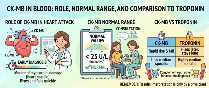
CK-MB appears in a lab report alongside troponin — and many patients wonder why two markers are ordered at the same time. The answer lies in their different diagnostic "windows": troponin remains elevated for up to 14 days, while CK-MB normalizes within 48–72 hours. This characteristic makes CK-MB indispensable for detecting reinfarction and monitoring the effectiveness of reperfusion therapy.
What CK-MB Is and How CK Isoenzymes Work
CK (creatine kinase, creatine phosphokinase) is an enzyme that catalyzes the reversible phosphorylation of creatine to produce ATP. It supplies rapid energy to cells with high energy demand — primarily muscle and nerve cells.
CK consists of two subunits — M (muscle) and B (brain) — and exists in three isoenzyme forms:
| Isoenzyme | Subunits | Primary location | Normal proportion |
|---|---|---|---|
| CK-MM | M + M | Skeletal muscle | 95–97% |
| CK-MB | M + B | Myocardium (15–25% of cardiac CK) | 3–5% |
| CK-BB | B + B | Brain, smooth muscle | < 1% |
CK-MB is expressed predominantly in cardiomyocytes: in heart muscle it accounts for 15–25% of total CK. In skeletal muscle, CK-MB comprises less than 3% — this concentration gradient is the biochemical basis of the marker's cardiac specificity.
When cardiomyocytes undergo necrosis or severe injury, CK-MB enters the bloodstream and becomes measurable. The mechanism parallels that of troponin, but CK-MB is released more rapidly and clears from the circulation faster — the difference in "memory" is what gives each marker its distinct clinical role.
Normal CK-MB Levels
Laboratories use two measurement approaches for CK-MB — activity-based and mass-based. They reflect different analytical dimensions and differ in sensitivity.
CK-MB activity (catalytic enzyme activity):
| Group | Normal |
|---|---|
| Adult men | < 25 U/L |
| Adult women | < 25 U/L |
| Children | < 30 U/L |
CK-MB mass (immunochemical determination — more sensitive):
| Group | Normal |
|---|---|
| Adults | < 5.0–6.0 µg/L (ng/mL) |
CK-MB/total CK ratio — an additional diagnostic criterion:
- < 3% — normal ratio; total CK elevation is most likely skeletal in origin
- 3–6% — borderline
- ≥ 6% — suggests cardiac origin when both CK-MB and total CK are elevated
The ratio is particularly useful in massive skeletal muscle injury (rhabdomyolysis, extreme exercise), where very high total CK "dilutes" the apparent MB fraction percentage.
CK-MB Kinetics in Acute Myocardial Infarction
The temporal profile of CK-MB in myocardial infarction is highly predictable — and this predictability is one of the marker's primary clinical strengths.
| Time from symptom onset | CK-MB activity |
|---|---|
| 0–3 hours | Normal or borderline |
| 3–6 hours | Rising in most patients |
| 12–24 hours | Peak activity |
| 48–72 hours | Normalization |
With successful reperfusion (thrombolysis or PCI), the CK-MB curve changes characteristically: the peak arrives earlier (6–12 hours) and is higher — the "reperfusion peak" — reflecting rapid washout of the marker from the re-opened segment. Early, high CK-MB peak followed by rapid decline after reperfusion is an indirect laboratory sign of successful blood flow restoration.
Collection protocol for MI monitoring:
- First draw: at presentation (hour 0)
- Repeat at 3–4 hours
- Again at 6–8 hours if diagnosis remains uncertain
- Immediately when reinfarction is suspected — at onset of new symptoms
No special preparation is required — fasting status does not affect CK-MB. In acute settings the test is ordered immediately, without any preparation.
Causes of Elevated CK-MB
A rise in CK-MB is almost always a sign of myocardial injury or — less commonly — severe skeletal muscle damage.
Cardiac causes:
| Condition | Degree of elevation | Characteristic features |
|---|---|---|
| Acute myocardial infarction | Significant (5–10× ULN or higher) | Acute onset; chest pain; ECG changes |
| Unstable angina | Normal or mild | Usually normal — key distinction from MI |
| Myocarditis | Moderate | Viral prodrome; chest discomfort |
| Cardioversion / defibrillation | Mild–moderate | Transient rise after procedure |
| Cardiac surgery | Significant | Expected postoperative pattern |
| Cardiac contusion | Moderate–significant | Chest trauma |
| Prolonged tachyarrhythmias | Mild | During sustained AF or VT episodes |
Non-cardiac causes (significant skeletal muscle damage releases CK-MB from its small muscle fraction):
| Condition | Mechanism | How to distinguish |
|---|---|---|
| Rhabdomyolysis | Massive skeletal muscle cell death | CK-MB/CK ratio < 6%; very high total CK |
| Intense training, marathon | Muscle stress | Transient; no infarction symptoms |
| Muscular dystrophies (regeneration) | Regenerating muscle expresses CK-MB | Chronic elevation; chronic disease context |
| Hypothyroidism | Reduced CK clearance | Elevated TSH; multiple enzyme elevations |
| Severe kidney failure | Reduced clearance | Changes in CBC and other markers |
| Pulmonary embolism | Right ventricular pressure overload | Clinical picture of PE; ECG |
C-reactive protein in acute MI rises alongside CK-MB — but later (peaking at 48–72 hours) and persisting longer. Interpreting these markers together helps distinguish the early infarction phase from the subsequent inflammatory response to myocardial necrosis.
CK-MB vs Troponin: The Fundamental Difference
With the advent of high-sensitivity troponin, the role of CK-MB in diagnosis has evolved — but not disappeared. Understanding their differences determines when each marker is needed.
| Characteristic | CK-MB | Troponin (hsTn) |
|---|---|---|
| Onset of rise | 3–6 hours | 1–3 hours (hsTn) |
| Peak | 12–24 hours | 12–24 hours |
| Normalization | 48–72 hours | 7–14 days |
| Sensitivity | Lower | Substantially higher |
| Cardiac specificity | High, but lower than troponin | Very high (cTnI) |
| Primary role today | Reinfarction diagnosis; reperfusion monitoring | Primary MI diagnosis |
| Detection of small MI | Misses some | Detects |
Key conclusion: for primary infarction diagnosis, troponin is preferred — it is more precise and responds earlier. But when troponin is already elevated (where it stays for up to 2 weeks), CK-MB is the only marker that can detect a new infarction: a fresh CK-MB rise on a background of persistently elevated troponin is the diagnostic sign of reinfarction.
CK-MB in Reinfarction and Reperfusion Monitoring
These are the two main clinical niches of CK-MB in modern cardiology — and both remain relevant despite widespread adoption of high-sensitivity troponin.
Reinfarction diagnosis. After a first infarction, troponin remains elevated for up to 14 days. If the patient develops new chest pain during this period — how do you distinguish reinfarction from post-infarction chest discomfort? Troponin is no longer useful as a dynamic marker. CK-MB has already normalized — and a new CK-MB rise of more than 20% from the previous value on repeat sampling 3–6 hours later is the diagnostic criterion for reinfarction under current guidelines.
Reperfusion monitoring. After successful thrombolysis or PCI, washout of markers from reperfused myocardium accelerates: an early CK-MB peak (< 12 hours), high and rapidly declining, is a sign of successful reperfusion. A delayed, flat peak without early decline may suggest incomplete reperfusion. This "non-invasive reperfusion index" provides a laboratory surrogate for procedural success.
Additionally, the absolute height of the CK-MB peak is used to roughly estimate infarct size — a higher peak correlates with a larger necrotic zone and worse prognosis.
When Elevated CK-MB Requires Medical Attention
CK-MB is not a routine screening test. It is ordered for specific clinical indications and almost exclusively in the context of acute cardiac presentations.
Call emergency services immediately for any CK-MB elevation combined with:
- Chest pain or pressure, breathlessness, or palpitations
- ECG changes — regardless of CK-MB level
- A new CK-MB rise in a patient who has already had a myocardial infarction
Scheduled cardiology consultation when:
- Moderate CK-MB elevation without clinical acute infarction — to exclude myocarditis, cardiac contusion, or other causes
- Chronically elevated CK-MB with a normal CK-MB/total CK ratio — possible myopathy or hypothyroidism
Important: isolated moderate CK-MB elevation with normal troponin and normal ECG requires repeat measurement 3–6 hours later — dynamics matter more than a single value. A stable level without rise substantially reduces the probability of acute infarction.
This article is for informational purposes only and does not replace professional medical advice. Call emergency services immediately if you experience chest pain.
For informational purposes only
This article is for informational purposes only and does not constitute medical advice, diagnosis, or treatment. Please consult a healthcare professional for medical guidance.