D-Dimer Blood Test: Normal Levels, Causes and Significance
Reviewed by the LabReadAI medical team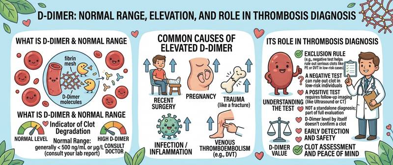
D-dimer is one of the few laboratory markers used primarily to rule out a diagnosis rather than confirm one. A normal D-dimer combined with low clinical probability excludes pulmonary embolism and deep vein thrombosis with more than 99% accuracy — without CT angiography or ultrasound. This is precisely what makes it indispensable in emergency medicine. Let's break down where D-dimer comes from, why it rises, and how to interpret results correctly.
What D-Dimer Is and How It Forms
D-dimer is a small protein fragment produced when a fibrin clot is broken down. To understand its origin, the coagulation cycle must be understood.
When a blood vessel is damaged, the coagulation cascade is activated: thrombin converts fibrinogen into fibrin, fibrin strands form a three-dimensional clot network, which is then stabilized by factor XIII. In parallel, the fibrinolytic system activates — plasmin degrades the fibrin clot to prevent excessive thrombosis. Breakdown of cross-linked (stabilized) fibrin produces characteristic fragments — D-dimers: each molecule contains two "D" domains from two adjacent fibrin molecules connected by cross-links.
D-dimer is simultaneously a marker of two processes: thrombosis (no clot = no fibrin = no D-dimer) and fibrinolysis (no clot breakdown = no D-dimer release). An elevated D-dimer therefore signals an active thrombotic process — somewhere in the body, clots are both forming and being dissolved.
In deep vein thrombosis and pulmonary embolism, this process reaches clinically significant proportions. This is why D-dimer is the first laboratory test ordered when venous thromboembolism is suspected.
Normal D-Dimer Levels
D-dimer reference values depend on the assay method and units used — it is one of the least standardized markers in laboratory diagnostics.
Standard adult reference range:
| Units | Normal |
|---|---|
| µg/mL FEU (fibrinogen equivalent units) | < 0.5 µg/mL |
| ng/mL FEU | < 500 ng/mL |
| µg/mL DDU (D-dimer units) | < 0.25 µg/mL |
| ng/mL DDU | < 250 ng/mL |
Important: FEU = 2 × DDU. Different laboratories use different units, and the same value of 500 ng/mL may be normal in one unit system and pathological in another. Always compare against the reference range printed on your specific laboratory report.
Age-adjusted threshold (for patients > 50 years): Clinical practice uses the formula age × 10 ng/mL (FEU). For a 70-year-old patient, the PE exclusion threshold rises to 700 ng/mL instead of the standard 500 ng/mL. This increases specificity without sacrificing sensitivity in older patients.
During pregnancy, D-dimer physiologically rises trimester by trimester:
| Trimester | Approximate normal range (ng/mL FEU) |
|---|---|
| 1st trimester | < 700–800 |
| 2nd trimester | < 1000–1200 |
| 3rd trimester | < 1500–2000 |
Pregnancy is a critical interpretive challenge: physiological elevation renders the standard 500 ng/mL threshold essentially meaningless. When PE is suspected in a pregnant patient, trimester-specific thresholds or supplementary imaging are used.
How to Prepare for a D-Dimer Blood Test
D-dimer is measured in citrate plasma (blue-top sodium citrate tube). No special preparation is required, but several factors influence results.
- No fasting required — the test can be drawn at any time
- Immediate laboratory delivery: citrate plasma is unstable at room temperature beyond 2–4 hours
- Correct tube fill: under-filling the citrate tube alters the anticoagulant-to-blood ratio and distorts results
- Disclose anticoagulant therapy (warfarin, heparin, DOACs) — these reduce D-dimer by suppressing clot formation
- A coagulation panel is ordered concurrently when a thrombotic event is suspected — for complete hemostatic assessment
- D-dimer is not a routine screening test: it is ordered for specific clinical indications, not preventively
Causes of Elevated D-Dimer
Elevated D-dimer is a non-specific finding: it signals active fibrinolysis somewhere in the body but does not indicate its location or cause.
| Cause | Degree of elevation | Characteristic features |
|---|---|---|
| Deep vein thrombosis | Moderate–significant | Swelling, pain, erythema of limb |
| Pulmonary embolism | Significant | Dyspnea, pleuritic chest pain, tachycardia |
| DIC syndrome | Very high | Pancytopenia; simultaneous bleeding + thrombosis |
| Myocardial infarction | Moderate | ECG changes; elevated troponin |
| Stroke | Moderate | Neurological deficits |
| Malignancies | Moderate–significant | Chronic elevation |
| Sepsis and severe infection | Significant | Systemic infection picture |
| Pregnancy | Physiological | Rises through trimesters |
| Postoperative period | Moderate | Expected within first 1–2 weeks |
| Preeclampsia | Significant | Hypertension; proteinuria; edema |
| Chronic heart failure | Mild–moderate | Correlates with severity |
| Trauma and fractures | Moderate | Temporal link to injury |
| Antiphospholipid syndrome | Moderate | Recurrent thrombosis; pregnancy losses |
C-reactive protein in thrombotic events rises in parallel — through the inflammatory response to tissue necrosis. In myocardial infarction, D-dimer rises modestly within the first hours while CRP reaches its peak at 24–48 hours.
DIC (disseminated intravascular coagulation) is the most dangerous cause of sharply elevated D-dimer: massive systemic coagulation activation with simultaneous thrombosis and bleeding. D-dimer > 5000–10,000 ng/mL combined with thrombocytopenia and prolonged PT — diagnostic criteria for DIC.
The Primary Role of D-Dimer: Excluding PE and DVT
D-dimer is above all an exclusion tool, not a confirmation test. This is its defining characteristic, distinguishing it from most laboratory markers.
The clinical logic:
When PE or DVT is suspected, the physician first estimates the pre-test clinical probability using validated scoring systems — the Wells Score or the Geneva Score. The result stratifies patients:
Low or intermediate clinical probability:
- D-dimer < 500 ng/mL (or below the age-adjusted threshold) → PE/DVT excluded with > 99% probability → CT angiography is not needed
- D-dimer ≥ 500 ng/mL → imaging is required (CT pulmonary angiography for PE; compression ultrasound for DVT)
High clinical probability:
- D-dimer is not used — proceed directly to imaging regardless of the result
This algorithm saves countless patients from unnecessary radiation exposure from CT. But it only works when applied correctly — only in patients with low or intermediate pre-test probability.
What D-dimer does NOT do:
- Does not identify the location of a clot
- Does not confirm PE when elevated (specificity is only 40–50%)
- Is not used to monitor anticoagulant treatment
- Is not a screening test in asymptomatic patients
False Results: When D-Dimer Misleads
Falsely elevated D-dimer (elevated result, but no thrombosis):
- Pregnancy — physiologically high in all pregnant women
- Older age — chronic mild elevation without thrombosis occurs in > 50% of people over 80
- Any inflammation, infection, or malignancy
- Postoperative and post-trauma periods
- Liver disease — impaired clearance of fibrin degradation products
- Rheumatological conditions (SLE, rheumatoid arthritis)
This is precisely why a positive (high) D-dimer alone is not a diagnosis of thrombosis.
Falsely normal D-dimer (thrombosis present but D-dimer is normal):
- Very early-stage thrombosis (< 6–8 hours from onset) — fibrinolysis not yet significantly activated
- Small isolated clot (e.g., small distal DVT)
- Anticoagulant therapy already started — reduces D-dimer generation
- Chronic thrombosis without active fibrinolysis
A normal D-dimer in the setting of high clinical probability for PE is not grounds for excluding the diagnosis — imaging is still required.
When D-Dimer Requires Medical Attention
D-dimer is never interpreted in isolation — only in clinical context alongside symptoms and pre-test probability.
Call emergency services immediately if D-dimer is elevated combined with:
- Sudden breathlessness, chest pain, or hemoptysis — possible PE
- Swelling, pain, and redness of one leg — possible DVT
- Neurological symptoms or altered consciousness — possible stroke
- Simultaneous bleeding and thrombosis — possible DIC
Scheduled investigation for moderately elevated D-dimer without acute symptoms:
- Oncological workup for persistently elevated D-dimer without an identifiable cause
- Antiphospholipid syndrome testing for recurrent thromboses
- Hemostatic system assessment when D-dimer is repeatedly elevated
After successful thrombosis treatment, a persistently elevated D-dimer at 3–6 months is an independent recurrence risk factor. When deciding whether to discontinue anticoagulation after a first unprovoked VTE, D-dimer measured one month after stopping therapy is incorporated into decision algorithms.
This article is for informational purposes only and does not replace professional medical advice. Seek immediate medical care if symptoms of thrombosis or pulmonary embolism are present.
For informational purposes only
This article is for informational purposes only and does not constitute medical advice, diagnosis, or treatment. Please consult a healthcare professional for medical guidance.