Coagulation Test: What's Included, Normal Values and Results
Reviewed by the LabReadAI medical team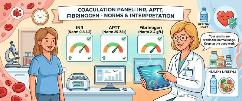
Blood must be able to do two opposite things at once: stop bleeding instantly when a vessel is damaged — while never clotting inside intact vessels where a clot should not form. This fragile equilibrium is maintained by the haemostasis system, and when it fails in either direction — excessive clotting or its deficiency — the consequences can be life-threatening. A coagulation test is a group of laboratory assays that evaluate the clotting system from multiple angles simultaneously. This article covers what the panel includes, how to prepare, what normal values look like, and what each deviation means.
What Is a Coagulation Test and How Does the Clotting Cascade Work?
Blood coagulation is a multi-step biochemical cascade in which clotting factors are sequentially activated until a firm fibrin clot forms and seals the site of injury. The process is divided into two converging pathways.
The extrinsic pathway is triggered when tissue is damaged outside the vessel wall: tissue factor (factor III) meets factor VII and initiates the cascade. This pathway is fast — and it is what prothrombin time measures.
The intrinsic pathway activates when blood contacts damaged vessel wall from inside: it is slower and more complex, initiated through factors XII, XI, IX, and VIII. APTT assesses this pathway.
Both pathways converge on factor X, after which thrombin is generated along the common pathway, and soluble fibrinogen is converted into insoluble fibrin. Simultaneously, the anticoagulant system — antithrombin, proteins C and S — prevents the clot from propagating beyond the injury site. A third system — fibrinolysis — dissolves the clot once healing is complete.
A coagulation panel evaluates all three elements: the speed of clot formation, the status of individual factors, and fibrinolytic activity.
When Is a Coagulation Test Ordered?
Indications range from emergencies to routine pre-procedure screening. A coagulation panel is mandatory before any surgical procedure or invasive intervention — unrecognised clotting defects can turn intraoperative bleeding into a catastrophe.
Symptomatic indications:
- Unexplained bleeding: nosebleeds, gum bleeding, uterine, or gastrointestinal haemorrhage
- Bruising without trauma (petechiae, ecchymoses)
- Prolonged bleeding after minor cuts or dental extractions
- Suspected deep vein thrombosis or pulmonary embolism
- Unexplained leg pain with swelling — possible thrombosis
Routine and monitoring indications:
- Pre-operative preparation — mandatory before any surgery
- Pregnancy — at booking, each trimester, and before delivery
- Monitoring anticoagulant therapy (warfarin, heparin, direct oral anticoagulants)
- Liver disease — the liver synthesises most clotting factors
- Diagnosis of DIC in severe infection, sepsis, or shock
- Malignancy — cancer substantially elevates thrombotic risk
- Systemic autoimmune diseases: antiphospholipid syndrome, lupus
How to Prepare and Get Tested
Blood for a coagulation panel is drawn into a tube containing sodium citrate, which binds calcium and halts clotting until analysis begins. Sample collection technique matters here more than in almost any other blood test — errors produce false results with striking frequency.
Preparation guidelines:
- Blood must be drawn strictly fasting — at least 8–12 hours after the last meal. A fatty meal beforehand causes lipaemia that interferes with optical detection methods.
- Avoid alcohol for 24 hours — it impairs hepatic synthesis of clotting factors.
- Do not smoke for 30 minutes before the draw: nicotine causes transient platelet activation.
- Inform your doctor of all medications. Anticoagulants (warfarin, rivaroxaban, heparin), antiplatelets (aspirin, clopidogrel), oral contraceptives, antibiotics, and NSAIDs all significantly affect results.
- Do not stop anticoagulant therapy before the test without medical instruction — this is dangerous.
- The tube must not be shaken or chilled: haemolysis of the sample markedly shortens the prothrombin time and invalidates the entire panel.
- When monitoring warfarin therapy — always test at the same time of day: INR fluctuates slightly across the day.
A critical technical point: the ratio of blood to citrate in the tube must be exactly 9:1. An underfilled tube disrupts this ratio, and results — particularly APTT and INR — will be falsely prolonged. The laboratory is obligated to reject an incorrectly filled tube.
What the Panel Includes: PT, INR, APTT, Fibrinogen and D-dimer
The composition of the panel depends on the clinical question — from a minimal screen to a full thrombophilia workup.
Prothrombin time (PT) and prothrombin index (PTI) assess the extrinsic and common pathways. PT is measured in seconds; normal in adults is 11–16 seconds. PTI is the ratio of normal PT to the patient's PT, expressed as a percentage; normal is 80–120%. Prolonged PT and reduced PTI indicate impairment of the extrinsic pathway: deficiency of factors VII, X, V, II, or hepatic failure.
INR (International Normalised Ratio) is a standardised expression of the PT result that allows comparison across laboratories. Normal in healthy individuals: 0.85–1.15. INR is the primary marker for warfarin therapy monitoring: the therapeutic range for thrombosis treatment is 2.0–3.0. A value above 3.5 without a therapeutic reason signals bleeding risk; below 0.8 may indicate hypercoagulability.
APTT (activated partial thromboplastin time) evaluates the intrinsic pathway. Normal: 25–37 seconds. APTT is prolonged by deficiencies of factors VIII, IX, XI, and XII — including haemophilia A and B, antiphospholipid syndrome, and heparin therapy. A shortened APTT points to hypercoagulability.
Thrombin time (TT) evaluates the final step: the conversion of fibrinogen to fibrin by thrombin. Normal: 14–21 seconds. Prolongation indicates deficiency or structural abnormality of fibrinogen, or the effect of direct thrombin inhibitors (dabigatran).
Fibrinogen is the protein precursor to fibrin, synthesised in the liver. Normal: 2.0–4.0 g/L. Fibrinogen is an acute-phase protein: it rises sharply with inflammation, infection, and trauma. Chronically elevated fibrinogen is an independent cardiovascular risk factor. A level below 1.5 g/L reflects consumption in DIC or severe hepatic failure.
D-dimer is the breakdown product of fibrin — a marker of active clot formation and fibrinolysis. It is the only coagulation panel indicator that measures not clotting potential but its real-time activation. For the full clinical significance of this marker and its role in thrombosis diagnosis, see D-dimer.
Antithrombin III (AT III) is the primary physiological inhibitor of clotting. Normal: 80–120%. Reduced levels indicate inherited or acquired deficiency — with a sharply elevated thrombotic risk. Ordered when thrombophilia is suspected.
Platelets are evaluated alongside plasma factors because primary haemostasis is the platelet plug. A fall in the count or function of platelets with normal plasma factors is itself an independent cause of bleeding.
Coagulation Test Normal Values: Summary Table
Reference ranges depend on the analytical method and laboratory equipment — always check the values on your own report.
| Parameter | Adults | Pregnancy (3rd trimester) |
|---|---|---|
| PT (sec) | 11–16 | 9.5–13.5 |
| INR | 0.85–1.15 | 0.80–1.20 |
| PTI (%) | 80–120 | 90–130 |
| APTT (sec) | 25–37 | 17–30 |
| Thrombin time (sec) | 14–21 | 14–20 |
| Fibrinogen (g/L) | 2.0–4.0 | 3.5–6.5 |
| D-dimer (µg/L FEU) | < 500 | < 1000 (I) / < 1500 (II) / < 2000 (III) |
| Antithrombin III (%) | 80–120 | 85–110 |
Pregnancy physiologically shifts the coagulation system toward hypercoagulability — a protective mechanism against blood loss during delivery. APTT shortens, fibrinogen and D-dimer rise, and PT is slightly shortened. D-dimer norms in pregnancy are substantially higher than in non-pregnant adults, with a separate reference range for each trimester. Applying standard non-pregnant cut-offs to pregnant women leads to significant overdiagnosis of thrombosis.
Interpreting Deviations: Hypercoagulability and Hypocoagulability
A doctor reads the coagulation panel as an interconnected system of markers, not as isolated numbers. The pattern of combined changes points to the specific disorder.
Prolonged PT/INR with normal APTT — extrinsic pathway impairment: factor VII deficiency (the shortest-lived clotting factor, the first to fall in liver disease), start of warfarin therapy, or early DIC.
Prolonged APTT with normal PT — intrinsic pathway impairment: haemophilia A (factor VIII deficiency) or B (factor IX deficiency), von Willebrand disease, heparin therapy, antiphospholipid syndrome.
Both PT and APTT prolonged — common pathway impairment or multiple factor deficiency: severe hepatic failure, hypocoagulable DIC, vitamin K deficiency, massive transfusion.
DIC (disseminated intravascular coagulation) — one of the most dangerous haemostatic emergencies: initially, widespread thrombosis occurs (hypercoagulable phase: short APTT and PT, high D-dimer), then clotting factors and platelets are consumed — giving way to profound hypocoagulability with uncontrollable bleeding. DIC develops in severe sepsis, obstetric catastrophes, major trauma, and oncological complications.
Hypercoagulability — shortened APTT and/or PT, elevated fibrinogen, rising D-dimer — signals elevated thrombotic risk. Clinically significant patterns: antiphospholipid syndrome (paradoxically prolonged APTT combined with thrombotic episodes), thrombophilia (AT III deficiency, protein C or S deficiency, factor V Leiden), and malignancy. The thrombotic risks associated with hypercoagulable states are discussed in detail in the article deep vein thrombosis.
Isolated D-dimer elevation with normal PT and APTT is not a clotting disorder — it is a marker of an existing clot already formed and being dissolved. D-dimer is used to rule out pulmonary embolism in clinical decision pathways: a negative result in a low-probability patient avoids the need for CT pulmonary angiography. The diagnostic approach to suspected embolism is covered in pulmonary embolism.
When Coagulation Test Results Require Urgent Medical Attention
Most moderate coagulation abnormalities call for a scheduled appointment with a GP, haematologist, or obstetrician. But some situations are medical emergencies:
- INR above 5.0 in a patient on warfarin — risk of spontaneous haemorrhage
- APTT above 100 seconds or PT above 30 seconds without anticoagulant therapy
- Sharply elevated D-dimer with leg pain and swelling plus breathlessness — possible PE
- Fibrinogen below 1.0 g/L — imminent uncontrollable bleeding
- Simultaneous prolongation of PT and APTT, rising D-dimer, falling platelets — DIC
- Any coagulation abnormality in a pregnant woman with symptoms — immediate evaluation
- Heavy post-operative bleeding with a previously normal pre-operative panel
- Newly discovered prolonged APTT in a child with a bleeding history — rule out haemophilia
A special case: antiphospholipid syndrome. APTT is paradoxically prolonged in the test tube, while in the body the patient has hypercoagulability. Patients with this pattern and a history of thrombosis need rheumatology referral and antiphospholipid antibody testing.
Conclusion
A coagulation panel is not a single assay but a carefully designed set of markers, each illuminating a different component of the haemostatic system. PT and INR evaluate the extrinsic pathway; APTT the intrinsic; fibrinogen the raw material for the clot; D-dimer the evidence of its formation and breakdown. It is the combination of results — not any single value — that points to the nature of the disorder. Proper preparation — fasting, no alcohol, a full medication list, and a correctly filled collection tube — ensures reliable results. When abnormalities are found, particularly alongside clinical symptoms, do not attempt self-interpretation: disorders of haemostasis require full clinical context.
This content is for informational purposes only and does not replace professional medical advice.
For informational purposes only
This article is for informational purposes only and does not constitute medical advice, diagnosis, or treatment. Please consult a healthcare professional for medical guidance.