HDL Cholesterol: Normal Levels, Causes and How to Raise It
Reviewed by the LabReadAI medical team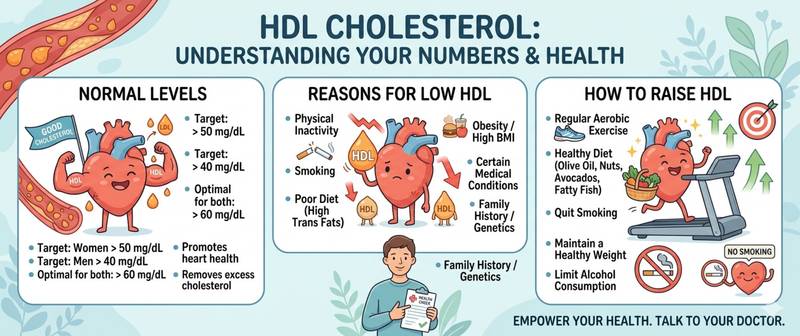
HDL is called "good cholesterol" — and while that is a fair label, the full picture is considerably more nuanced. High-density lipoproteins are not merely harmless compared to LDL: they actively protect arteries by collecting excess cholesterol from vessel walls and transporting it back to the liver. Low HDL is an independent cardiovascular risk factor — separate from LDL levels. Let's break down what makes HDL protective and why a low reading warrants attention.
What HDL Is and How Reverse Cholesterol Transport Works
HDL (high-density lipoproteins) are the smallest and densest of all lipoproteins. Unlike LDL, which carries cholesterol from the liver to peripheral tissues, HDL operates in the opposite direction: it collects excess cholesterol from peripheral tissues and arterial walls and delivers it back to the liver for processing and biliary excretion. This process is called reverse cholesterol transport and is the primary anti-atherogenic mechanism of HDL.
Beyond transport, HDL has several additional protective properties: it inhibits LDL oxidation (reducing the most atherogenic oxidized forms), suppresses endothelial inflammation, participates in fibrinolysis, and exerts antithrombotic effects.
HDL's key protein is apolipoprotein A-I (apo-A-I): it initiates cholesterol uptake from cells via the ABCA1 transporter and determines the particle's functional activity. Apo-A-I levels actually predict cardiovascular risk better than HDL concentration — but standard HDL measurement remains the routine clinical tool.
HDL is a component of the lipid panel, and its interpretation always proceeds alongside LDL, triglycerides, and total cholesterol.
HDL Normal Ranges: Sex Matters
Unlike most biochemical markers, HDL reference values differ substantially between men and women: women have on average 0.3–0.5 mmol/L higher HDL than men of the same age. This is one reason why women before menopause carry lower cardiovascular risk at equal LDL levels.
| Group | Optimal HDL | Borderline | Low (risk factor) |
|---|---|---|---|
| Men | > 1.4 mmol/L | 1.0–1.4 mmol/L | < 1.0 mmol/L |
| Women | > 1.7 mmol/L | 1.2–1.7 mmol/L | < 1.2 mmol/L |
Clinically relevant thresholds (ESC/EAS):
- < 1.0 mmol/L in men and < 1.2 mmol/L in women — independent cardiovascular risk factor
- Very high HDL (> 2.5–3.0 mmol/L) — paradoxically, this can also be associated with increased risk (dysfunctional HDL)
With age, HDL in men changes very little, while in women it falls after menopause — parallel to the drop in estrogen, which stimulates apo-A-I synthesis.
During pregnancy, HDL physiologically rises in the second trimester and falls toward the third — a normal trajectory requiring no intervention.
How to Prepare for an HDL Blood Test
HDL is measured in serum as a component of the lipid panel. Preparation requirements are standard across the full lipid profile.
- Strictly fasting: at least 12 hours without food. HDL is only modestly affected by eating, but fasting is required for the panel as a whole and for accurate Friedewald LDL calculation
- No alcohol for 48–72 hours: alcohol transiently raises HDL — giving a falsely optimistic result
- Stable diet for 2–3 weeks: dietary changes (especially adding omega-3s) alter HDL gradually — the result should reflect a typical baseline
- Acute illness lowers HDL by 10–30% — best to postpone testing 6–8 weeks after recovery
- For serial monitoring: same laboratory under identical conditions
HDL is a relatively stable marker — its analytical variability is lower than triglycerides. However, meaningful lifestyle changes require 8–12 weeks to register in test results.
Causes of Low HDL
Low HDL is far more common and clinically significant than elevated HDL. In most cases, reduction is driven by lifestyle and metabolic disorders — and those are precisely the targets of treatment.
| Cause | Mechanism | Characteristic features |
|---|---|---|
| Metabolic syndrome / insulin resistance | Accelerated HDL catabolism; reduced apo-A-I | High TG + low HDL + abdominal obesity |
| Type 2 diabetes | Glycation of apo-A-I impairs HDL function | Diabetic dyslipidemia: TG↑, HDL↓, small dense LDL |
| Physical inactivity | Reduced apo-A-I synthesis | Persistently low HDL in sedentary individuals |
| Smoking | Oxidative HDL damage; reduced apo-A-I synthesis | Especially pronounced in men |
| Obesity (abdominal pattern) | Enhanced HDL catabolism via VLDL exchange | Combined with high TG |
| Trans fat consumption | Reduced apo-A-I synthesis; increased CETP activity | Chronic margarine and industrial food intake |
| Hypothyroidism | Reduced lipoprotein lipase activity | Combined with elevated LDL |
| Chronic kidney disease | HDL dysfunction; accelerated catabolism | Parallel TG elevation |
| Medications | Reduced synthesis or enhanced catabolism | Beta-blockers, thiazides, progestins, anabolic steroids |
| Genetic causes (hypoalphalipoproteinemia) | ABCA1, APOA1, LCAT gene mutations | Very low HDL < 0.5 mmol/L |
The combination of high triglycerides + low HDL — "atherogenic dyslipidemia" — is the hallmark pattern of metabolic syndrome and insulin resistance. These markers are mechanistically linked: VLDL particles (carrying triglycerides) and HDL exchange components in the blood; when VLDL is in excess, HDL becomes enriched with triglycerides, becomes a substrate for lipases, and is rapidly degraded.
Causes of High HDL
High HDL is traditionally viewed as straightforwardly favorable — and in most cases it is. However, extremely high values warrant attention.
Physiological and favorable causes:
- Regular physical activity — the most effective way to raise HDL
- Moderate alcohol consumption (1–2 units per day) — raises HDL by 5–10%
- Female sex before menopause — estrogenic stimulation of apo-A-I synthesis
- Genetic variants with high apo-A-I activity
Potentially problematic causes of very high HDL (> 2.5–3.0 mmol/L):
- CETP gene mutations (cholesteryl ester transfer protein) — HDL is high but dysfunctional: reverse cholesterol transport is impaired
- Chronic heavy alcohol use — HDL is elevated but this is a falsely reassuring marker; cardiovascular risk is not reduced
- Primary biliary cholangitis — disrupted cholesterol metabolism
- Hyperthyroidism — accelerated HDL synthesis and catabolism
The paradox of "too-high HDL": several large studies showed a J-shaped association — at HDL > 2.5–3.0 mmol/L, cardiovascular event risk and total mortality begin to rise. Proposed mechanism: at extreme HDL concentrations, particles may acquire pro-inflammatory properties. This is an active research area, but clinicians now pay attention to unexplainably elevated HDL.
HDL in the Context of the Full Lipid Profile
HDL cannot be meaningfully assessed in isolation — its protective role is fully apparent only alongside the other lipid markers.
HDL and LDL: atherogenicity index. The most informative integrated risk measure is the LDL/HDL ratio or atherogenicity coefficient = (Total cholesterol − HDL) / HDL. Optimal value < 3.0; high risk at > 4.0–5.0. At normal LDL but low HDL, the atherogenicity index remains high — and cardiovascular risk is not mitigated.
HDL and triglycerides: the reciprocal link. As explained above, high TG and low HDL are mechanistically linked — they reflect the same pathogenic process. Any intervention that reduces triglycerides (lifestyle, omega-3, fibrates) predictably raises HDL.
HDL and atherosclerosis: the causal link between HDL and atherosclerosis is less straightforward than for LDL. Mendelian randomization data show that genetically high HDL does not always reduce heart attack risk. This means that what matters is not just HDL concentration, but HDL functionality — the actual capacity to carry out reverse cholesterol transport. Dysfunctional HDL at normal concentrations may provide no protection. This explains the disappointing results of clinical trials of drugs that simply raised HDL levels without improving their function.
How to Raise HDL and When to See a Doctor
HDL is one of the most difficult lipid markers to raise pharmacologically. This is precisely why lifestyle modification plays the primary role.
Most effective non-pharmacological approaches:
Physical activity — the most evidence-backed way to raise HDL. Moderate-to-vigorous aerobic exercise (running, swimming, cycling) for 150 minutes per week raises HDL by 5–15% over 8–12 weeks. Resistance training provides a smaller but meaningful additional effect.
Smoking cessation — within weeks of stopping, HDL rises by 5–10%.
Weight loss — every 3 kg of fat lost raises HDL by approximately 0.04 mmol/L; a 10–15% weight reduction in obese individuals produces clinically meaningful results.
Dietary changes: eliminate trans fats (raising HDL by 3–7%); increase monounsaturated fats (olive oil, avocado); adequate omega-3 intake (modest HDL rise via TG reduction).
Moderate alcohol consumption — biochemically raises HDL, but recommending alcohol as a cardioprotective measure is clinically unacceptable given its harm to the liver, brain, cancer risk, and addiction potential.
Pharmacological approaches:
No existing drug has been shown to reduce cardiovascular risk purely by raising HDL — this is the central conclusion from clinical trials over the past 15 years.
Statins modestly raise HDL (by 5–10%), but their cardioprotective effect is primarily attributable to LDL reduction. Fibrates and niacin substantially raise HDL (by 15–35%) and lower TG, but have not reduced cardiovascular mortality in isolated use. Fibrates are prescribed for severe hypertriglyceridemia, not for HDL elevation as a standalone goal.
See a doctor when:
- HDL < 1.0 mmol/L (men) or < 1.2 mmol/L (women) on repeat testing
- Low HDL combined with high triglycerides and any LDL level — full cardiometabolic risk assessment needed
- Family history of premature heart attack with unexplainably low HDL — genetic causes should be excluded
For HDL in the extended lipid profile context, its longevity role and link to metabolic health, see the article extended lipid profile and longevity.
This article is for informational purposes only and does not replace professional medical advice. Consult a cardiologist or GP if your HDL level is outside the normal range.
For informational purposes only
This article is for informational purposes only and does not constitute medical advice, diagnosis, or treatment. Please consult a healthcare professional for medical guidance.