RDW Blood Test: Normal Levels, Causes and Interpretation
Reviewed by the LabReadAI medical team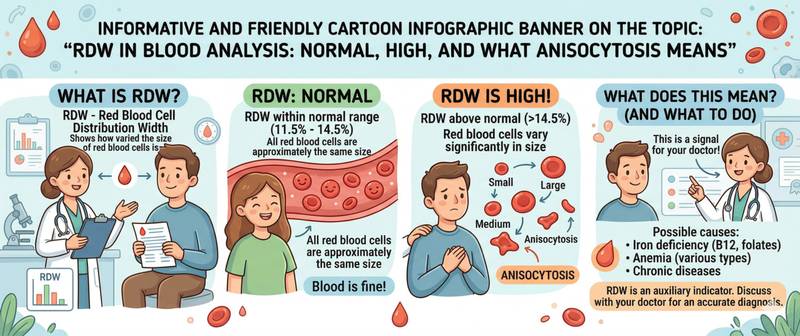
In a complete blood count report, RDW sits alongside hemoglobin and MCV, and most patients have no idea what to make of it. Yet it is one of the most informative markers for identifying the cause of anemia: it answers the question of how uniform your red blood cells are in size. Anisocytosis — size variation among red cells — is not just a laboratory term; it is a direct imprint of what is happening in the bone marrow. Let's break down how RDW works and why it should always be read alongside MCV.
What RDW Is and What It Measures
RDW — Red Cell Distribution Width — is a parameter reflecting the degree of heterogeneity in red blood cell volume. Simply put: how much individual red cells differ from one another in size.
An automated hematology analyzer measures the volume of every red cell in the sample and constructs a distribution curve. RDW is a statistical characteristic of that curve. The wider the spread, the higher the RDW — and the more pronounced the anisocytosis.
Two calculation variants may appear on a lab report:
RDW-CV (coefficient of variation) — expressed as a percentage. Reflects the relative spread of red cell volumes relative to the mean. The most widely used variant.
RDW-SD (standard deviation) — expressed in femtoliters (fL). Reflects the absolute spread of volumes and is more sensitive to a small subpopulation of abnormally large or small cells. Less dependent on mean cell volume.
RDW cannot be interpreted in isolation — its diagnostic value is fully realized only alongside MCV (mean corpuscular volume) and hemoglobin. Together these three parameters form a coordinate system for the differential diagnosis of anemia.
In a complete blood count, RDW is automatically reported by all modern analyzers as a standard parameter — no separate test order is required.
Normal RDW Values: RDW-CV and RDW-SD
Reference values depend on the calculation method. Always check which parameter — CV or SD — is reported on the specific lab form.
| Parameter | Normal in adults | Normal in infants under 6 months |
|---|---|---|
| RDW-CV | 11.5–14.5% | 14.9–18.7% |
| RDW-SD | 35–47 fL | 35–55 fL |
Key nuances:
Newborns and infants under 6 months have a physiologically higher RDW — this is normal, reflecting the transition from fetal to adult hemoglobin and the uneven maturation of red cells during this period.
Pregnancy modestly elevates RDW — particularly in the second and third trimesters, driven by hemodilution and functional iron deficiency.
After blood transfusion, RDW is transiently elevated — donor and patient red cells differ in size, broadening the distribution curve.
A minor elevation (RDW-CV 14.6–15.5%) with normal hemoglobin and no clinical symptoms is often a normal variant or early deficiency requiring monitoring but not immediate treatment.
How to Prepare for an RDW Test
RDW is calculated automatically as part of a standard complete blood count — no additional sample or separate test order is needed.
- Blood is drawn from a vein, fasting or 3–4 hours after a light meal — normal food intake does not significantly affect RDW
- The sample should be analyzed within 4–8 hours of collection: prolonged storage causes red cells to swell, which can artificially elevate RDW-SD
- Recent blood transfusion (< 4–6 weeks) produces a falsely elevated RDW — mixing of donor and patient red cells
- Starting treatment for deficiency anemia (iron, B12) transiently raises RDW: new young normal-size cells appear alongside existing abnormal ones — the spread temporarily increases
- For serial monitoring, use the same laboratory — different analyzers produce slightly varying results
Causes of High RDW (Anisocytosis)
An elevated RDW means red cells are heterogeneous: cells of noticeably different sizes are circulating simultaneously. This occurs when normal red cell maturation is disrupted or when cells from multiple "generations" with different characteristics enter the bloodstream together.
| Cause | Mechanism | Characteristic MCV combination |
|---|---|---|
| Iron deficiency anemia | Small hypochromic cells alongside normal ones | ↓ MCV + ↑ RDW |
| Vitamin B12 / folate deficiency | Large megaloblastic cells alongside normal ones | ↑ MCV + ↑ RDW |
| Mixed anemia (Fe + B12) | Simultaneously small and large cells | Normal MCV + ↑ RDW |
| Start of deficiency anemia treatment | New normal cells + existing abnormal cells | Transitional period |
| Hemolytic anemia | Red cell fragments + reticulocytes | ↑ or normal MCV + ↑ RDW |
| Blood transfusion | Donor and patient red cells coexisting | Variable |
| Sideroblastic anemia | Two populations: normal and hypochromic | ↑ RDW + normal/↓ MCV |
| Chronic liver disease | Disrupted red cell membrane, target cells | ↑ MCV + ↑ RDW |
| Myelodysplastic syndrome | Impaired bone marrow maturation | Variable, often ↑ RDW |
The most clinically significant combination is low MCV + high RDW with low hemoglobin: this is virtually pathognomonic of iron deficiency anemia and calls for immediate measurement of ferritin.
A separate point of interest: RDW as an inflammatory and cardiovascular risk marker. Research over the past 15 years consistently shows that elevated RDW (> 14.5%) with normal hemoglobin is independently associated with higher mortality in heart failure, sepsis, and cancer. The mechanism: chronic inflammation disrupts normal red cell maturation. This is not a diagnostic criterion but a prognostic signal worth noting.
Causes of Low RDW
RDW below 11.5% (CV) is rare and carries almost no independent clinical significance on its own.
Main situations:
- Hereditary spherocytosis — a genetic condition: all red cells are uniformly small and spherical; size variation is minimal. Characteristic combination: low or normal RDW + reduced MCV + elevated MCHC + hemolysis
- Thalassemia minor — uniformly small red cells without significant size spread. An important differentiating feature from iron deficiency: in thalassemia, RDW is normal or low; in iron deficiency, it is elevated
- Anemia of chronic disease in early stages — uniformly modest reduction without significant anisocytosis
RDW is precisely the key laboratory feature that separates iron deficiency anemia (RDW ↑) from thalassemia (RDW normal/↓) when both present with microcytosis.
RDW and MCV: Differential Diagnosis of Anemia
Combining RDW and MCV is the standard first-line algorithm for classifying anemia by mechanism. This is the Bessman classification, widely used in clinical practice.
| MCV | RDW | Most likely cause |
|---|---|---|
| ↓ Low (microcytosis) | ↑ High | Iron deficiency anemia |
| ↓ Low (microcytosis) | Normal | Thalassemia, anemia of chronic disease |
| Normal | ↑ High | Early Fe or B12 deficiency, mixed anemia, hemolysis |
| Normal | Normal | Anemia of chronic disease, acute blood loss |
| ↑ High (macrocytosis) | ↑ High | B12 or folate deficiency, hemolysis |
| ↑ High (macrocytosis) | Normal | Liver disease, hypothyroidism, aplastic anemia |
Practical takeaway: in a patient with microcytic anemia who needs to be distinguished between iron deficiency and thalassemia — the first step is RDW. An elevated RDW points to iron deficiency and prompts ferritin testing; a normal RDW favors thalassemia and prompts hemoglobin electrophoresis.
When MCV and hemoglobin are normal but RDW is isolated elevated — checking ferritin and early B12 markers makes sense: anisocytosis often precedes an overt fall in hemoglobin by months.
When RDW Abnormalities Require Medical Attention
An isolated elevated RDW with normal hemoglobin and no symptoms warrants scheduled investigation — not emergency action.
Scheduled visit to a doctor when:
- RDW-CV > 14.5% at any hemoglobin level — to identify the cause of anisocytosis
- High RDW combined with low MCV — rule out iron deficiency anemia (ferritin, iron, TIBC)
- High RDW combined with high MCV — rule out B12 and folate deficiency
- Persistently elevated RDW on multiple sequential tests — even with normal hemoglobin
Seek urgent care when:
- High RDW combined with anemia (hemoglobin below normal) and symptoms: breathlessness, weakness, pallor, tachycardia
- RDW-CV > 17–18% — marked anisocytosis requiring exclusion of serious pathology (hemolysis, myelodysplasia)
- Anemia with elevated RDW following recent chemotherapy or radiotherapy
After starting deficiency anemia treatment, a transient RDW rise over 1–2 weeks is expected (the "bimodal" distribution as new normal cells appear) — this is a sign of treatment response, not deterioration.
This article is for informational purposes only and does not replace professional medical advice. Consult a GP or hematologist if your RDW is outside the normal range.
For informational purposes only
This article is for informational purposes only and does not constitute medical advice, diagnosis, or treatment. Please consult a healthcare professional for medical guidance.