Ferritin in Blood: Normal Levels, Causes and Significance
Reviewed by the LabReadAI medical team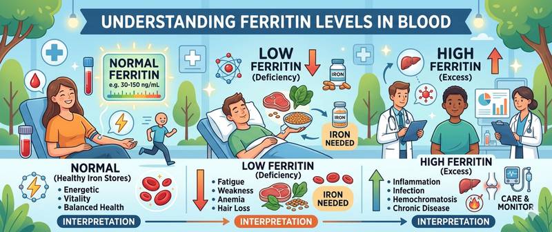
Ferritin is more than just an "iron test." It is a storage protein that reflects true iron reserves in the body far more accurately than serum iron itself. Serum iron can be normal even when reserves are completely depleted, while ferritin will reveal the deficiency weeks before anemia develops. This is precisely why ferritin is the first test ordered when iron deficiency is suspected and an essential component of a full iron status panel.
What Ferritin Is and What Role It Plays
Ferritin is an intracellular iron-storage protein: each ferritin molecule can hold up to 4,500 iron atoms in a non-toxic, biologically available form. It is present in virtually all cells but is most concentrated in hepatocytes, bone marrow macrophages, and reticuloendothelial cells.
In serum, ferritin circulates in small amounts — it "leaks" from cells in proportion to their iron content. This is why serum ferritin correlates with total body iron stores: 1 µg/L of serum ferritin ≈ 8–10 mg of stored iron.
Functions of ferritin:
- Iron storage — protects cells from toxic free iron (Fe²⁺), which generates reactive oxygen species via the Fenton reaction
- Erythropoiesis regulation — iron deficiency impairs hemoglobin synthesis and red cell production
- Acute phase protein — during inflammation, ferritin synthesis rises sharply regardless of iron stores; this is critically important for interpreting elevated results
A detailed clinical guide to interpreting ferritin across different clinical situations is available in ferritin: how to read your blood test.
Normal Ferritin Levels
Reference values depend substantially on sex and age. Women have significantly lower iron stores due to menstrual losses.
| Group | Normal ferritin (µg/L / ng/mL) |
|---|---|
| Newborns | 25–200 |
| Infants 1–5 months | 50–200 |
| Children 6 months – 15 years | 7–140 |
| Men 18–45 years | 20–250 |
| Men 45–65 years | 20–300 |
| Women 18–45 years (premenopausal) | 10–120 |
| Women postmenopause | 20–200 |
| Pregnant (1st trimester) | 10–90 |
| Pregnant (3rd trimester) | 5–50 |
Clinically relevant thresholds:
| Ferritin level (µg/L) | Interpretation |
|---|---|
| < 12–15 | Absolute iron deficiency |
| 12–30 | Depleted stores (pre-latent deficiency) |
| 30–100 | Normal (lower range) |
| 100–300 | Normal (mid to upper range) |
| > 300 (F) / > 400 (M) | Elevated — possible excess or inflammation |
| > 1000 | Significant elevation — active diagnostic workup required |
Key nuance: in patients with chronic inflammatory disease, a "normal" ferritin of 50–100 µg/L may conceal genuine iron deficiency — inflammation raises ferritin independently of iron stores. In the presence of active inflammation, ferritin < 30–50 µg/L is the diagnostically meaningful threshold for iron deficiency.
How to Prepare for a Ferritin Blood Test
Ferritin is a relatively stable marker, but several factors influence the result.
- Fasting for at least 8 hours is recommended — food has little direct effect on ferritin, but fasting is required when the full iron panel (including serum iron) is ordered simultaneously
- Stop iron supplements 5–7 days before the test — iron-containing medications transiently elevate ferritin
- Acute illness, infection, or active inflammation significantly raise ferritin as an acute phase protein — defer testing 6–8 weeks after resolution or interpret alongside CRP
- For serial monitoring: same laboratory each time
- Blood transfusion in the preceding 2–4 weeks distorts the result
Ferritin is always interpreted alongside other iron status markers: serum iron, TIBC (transferrin), transferrin saturation, and when needed — soluble transferrin receptor (sTfR).
Causes of Low Ferritin
Low ferritin is the most reliable marker of depleted iron stores. There are virtually no other causes of sub-normal ferritin.
| Cause | Mechanism | Characteristic features |
|---|---|---|
| Chronic blood loss (menstruation, GI bleeding) | Iron losses exceed intake | Most common cause in women |
| Inadequate dietary iron intake | Insufficient substrate for storage synthesis | Vegetarian and vegan diets; restrictive eating |
| Impaired iron absorption | Reduced duodenal absorption | Celiac disease, Crohn's disease, gastrectomy |
| Pregnancy and lactation | Increased iron demand | Physiological fall in 3rd trimester |
| Chronic iron deficiency anemia | Sustained deficiency with inadequate treatment | Low hemoglobin + low ferritin |
| Intense physical training | "Footstrike" hemolysis, myoglobinuria, sweat losses | Endurance athletes |
| Regular blood donation | Direct iron loss with each donation | Frequent donors |
In iron deficiency anemia, ferritin falls first — well before hemoglobin drops. The sequence of iron depletion: ferritin ↓ → transferrin saturation ↓ → MCHC ↓ → hemoglobin ↓. Ferritin is the only marker that catches iron deficiency before anemia develops.
Symptoms of iron depletion without anemia ("pre-latent deficiency"): chronic fatigue, difficulty concentrating, hair loss, restless legs syndrome, reduced exercise capacity. Many patients live for years with ferritin of 8–15 µg/L and normal hemoglobin, unable to explain why they feel chronically unwell.
Causes of High Ferritin (Hyperferritinemia)
Elevated ferritin is one of the most common "puzzling" biochemical findings: it can reflect iron accumulation, systemic inflammation, or serious disease.
| Cause | Ferritin level | Characteristic features |
|---|---|---|
| Inflammation and infection | Moderate (100–500 µg/L) | Elevated CRP; acute illness signs |
| Non-alcoholic fatty liver disease | Moderate–significant | Obesity; elevated ALT; insulin resistance |
| Alcoholic liver disease | Moderate–significant | AST/ALT > 2; elevated GGT |
| Hereditary hemochromatosis | Significant (> 500–1000 µg/L) | Transferrin saturation > 45%; HFE mutation |
| Malignancies | Variable, often > 1000 µg/L | Often unrelated to actual iron stores |
| Hemophagocytic lymphohistiocytosis (HLH) | Very high (> 10,000 µg/L) | Pancytopenia; fever; splenomegaly |
| Multiple blood transfusions | Significant | Transfusional iron overload |
| Chronic kidney disease | Moderate | Parallel erythropoiesis impairment |
| Metabolic syndrome | Mild–moderate | Obesity; high TG; hypertension |
| Hyperthyroidism | Mild | Falls with TSH normalization |
Hereditary hemochromatosis is the most important cause of significantly elevated ferritin that must not be missed. Iron accumulation in the liver, pancreas, heart, and joints — if untreated — leads to cirrhosis, diabetes, cardiomyopathy, and arthropathy. Screening marker: transferrin saturation > 45% combined with ferritin > 300–400 µg/L. Confirmed by genetic testing for the C282Y mutation in the HFE gene.
HLH (hemophagocytic lymphohistiocytosis) — a life-threatening condition: ferritin rises to 10,000–100,000 µg/L and is a diagnostic criterion. Every clinician should recognize this threshold.
Ferritin as an Iron Marker vs. an Inflammation Marker
The central diagnostic challenge with ferritin is that it simultaneously reflects two entirely different things: iron stores and the inflammatory response. This makes interpretation non-trivial.
In health: ferritin reflects iron stores — low = deficiency; high = excess.
In inflammation, infection, or malignancy: ferritin synthesis in the liver and macrophages is sharply upregulated as an acute phase response — independent of actual iron stores. This masks true iron deficiency:
Classic diagnostic trap: a patient with rheumatoid arthritis has ferritin of 80 µg/L — seemingly normal. But if CRP is 40 mg/L, the ferritin in the absence of inflammation would be around 15–20 µg/L — indicating significant iron deficiency. This is why ferritin must always be interpreted alongside CRP and the soluble transferrin receptor (sTfR) — the latter rises in iron deficiency and does not respond to inflammation.
Practical decision framework:
- Low ferritin + normal CRP → absolute iron deficiency; treat
- Normal/high ferritin + high CRP → possible occult deficiency; check sTfR
- High ferritin + normal CRP → iron overload or storage disease
When Ferritin Abnormalities Require Medical Attention
Scheduled visit to a doctor when:
- Ferritin < 20–30 µg/L — especially in women and with symptoms (fatigue, hair loss, exercise intolerance) — prescribe iron supplementation and investigate the cause
- Ferritin > 300 µg/L (women) or > 400 µg/L (men) without an obvious inflammatory condition — rule out hemochromatosis and liver disease
- Ferritin > 1000 µg/L — regardless of symptoms — active diagnostic workup required
Seek urgent care when:
- Ferritin > 5000–10,000 µg/L — rule out HLH and hematological malignancies
- Low ferritin combined with severe anemia (hemoglobin < 90 g/L)
- Rapidly rising ferritin in a patient with known disease — possible decompensation or malignant transformation
Ferritin is one of the key markers in biological age assessment and preventive health check-ups — see the article biological age by blood tests. For male-specific ferritin norms, causes of abnormal levels and correction strategies, see the article ferritin in men.
This article is for informational purposes only and does not replace professional medical advice. Consult a GP or hematologist if your ferritin level is outside the normal range.
For informational purposes only
This article is for informational purposes only and does not constitute medical advice, diagnosis, or treatment. Please consult a healthcare professional for medical guidance.