Transferrin Blood Test: Normal Range and Role in Anaemia Diagnosis
Reviewed by the LabReadAI medical team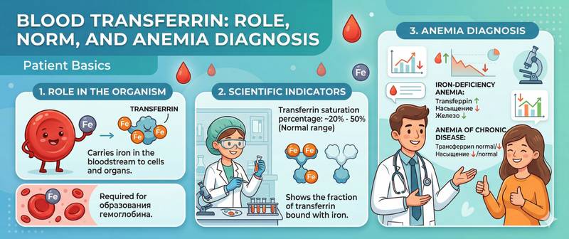
An iron panel often includes several markers at once — and transferrin occupies a unique position among them. It answers a question that ferritin alone cannot resolve: is the anaemia there because iron is genuinely insufficient, or because inflammation is locking it away in storage? This distinction fundamentally changes the treatment approach. Here's what transferrin measures, how to calculate its saturation, and how to read the results alongside other iron markers.
What Is Transferrin and Why Is It Measured
Transferrin is a transport protein synthesised by the liver. Its sole function is to carry iron through the bloodstream: capturing it at points of absorption (intestine) or release (breakdown of red cells in the spleen) and delivering it where it is needed — primarily to the bone marrow for haemoglobin synthesis.
A single transferrin molecule can bind two iron atoms. In health, only 20–45% of available sites are occupied — the rest remain free as reserve capacity. This free binding space is called the unsaturated iron-binding capacity (UIBC), and the total number of available sites is the total iron-binding capacity (TIBC).
The key calculated value is transferrin saturation (TSAT):
TSAT (%) = Serum iron / TIBC × 100
Normal TSAT: 20–45%. This percentage is the most informative single indicator of iron availability to the bone marrow in real time.
Transferrin is part of the standard iron panel alongside serum iron and ferritin. Together they provide a complete picture of iron metabolism — three different "levels": the current circulating level, the transport capacity, and the storage depot state.
Normal Transferrin and Saturation Values
| Marker | Normal Range |
|---|---|
| Transferrin | 2.0–3.6 g/L |
| TIBC | 45–75 µmol/L |
| UIBC | 25–56 µmol/L |
| TSAT (transferrin saturation) | 20–45% |
Women have slightly higher transferrin than men, reflecting lower baseline iron stores. During pregnancy, transferrin rises (to 4.0–4.5 g/L) — the body expands transport capacity for foetal needs.
Reference ranges vary slightly between laboratories. Always use the values on your specific report.
Transferrin in Iron-Deficiency Anaemia vs Anaemia of Chronic Disease
This is the primary diagnostic value of transferrin — and the main reason it is included in a comprehensive iron panel.
Two types of anaemia look similar on the surface (haemoglobin reduced, MCV may be low) but require fundamentally different treatment:
Iron-deficiency anaemia (IDA) — iron is genuinely insufficient. The body responds by producing more transferrin to "catch" every available iron atom. Transferrin is elevated, TIBC is elevated, ferritin is low, TSAT is low (< 20%). Treatment: iron supplementation.
Anaemia of chronic disease (ACD) — iron stores are adequate but inflammation "locks" iron in depots and prevents its use. During inflammation, the liver reduces transferrin synthesis. Transferrin is low, ferritin is elevated (it is an acute-phase protein), TSAT is low or normal. Treatment: treat the underlying inflammatory condition — iron supplementation is ineffective or harmful.
| Marker | IDA | ACD |
|---|---|---|
| Transferrin | ↑ elevated | ↓ low |
| Ferritin | ↓ low | ↑ elevated |
| TSAT | ↓ low | ↓ low or normal |
| Serum iron | ↓ low | ↓ low |
TSAT is reduced in both situations — which is exactly why TSAT alone is insufficient. Only the combined analysis of transferrin and ferritin allows the two conditions to be distinguished.
A combination can also exist: a patient with rheumatoid arthritis (chronic inflammation) may simultaneously have genuine iron deficiency — ferritin will be modestly elevated but transferrin also elevated, and TSAT markedly reduced.
Why Is Transferrin High?
Iron deficiency — the most common cause. Compensatory response: no iron → more carriers. The more severe the deficiency, the higher the transferrin.
Pregnancy — physiological rise, most pronounced in the second and third trimester.
Oral contraceptives — the oestrogen component stimulates transferrin synthesis in the liver.
Chronic blood loss (menorrhagia, gastrointestinal bleeding) — ongoing iron losses drive a compensatory transferrin rise.
Why Is Transferrin Low?
Chronic inflammation and infection — cytokines (IL-6, TNF-α) suppress hepatic transferrin synthesis. Classic ACD pattern.
Liver disease — in cirrhosis, hepatitis, and fatty liver disease, synthetic function declines. Transferrin falls alongside ALT, AST, and albumin.
Nephrotic syndrome — transferrin is lost with protein through the kidneys.
Malnutrition and malabsorption — insufficient substrate for protein synthesis.
Iron overload (haemochromatosis) — feedback mechanism: excess iron → fewer carriers. TSAT is markedly elevated (> 60–70%).
Hereditary atransferrinaemia — extremely rare genetic disorder with near-absent transferrin. Despite paradoxically overloaded depots, the bone marrow cannot access iron and severe anaemia develops.
How to Prepare for the Iron Panel Transferrin Test
Transferrin is measured as part of the iron panel. Blood is drawn fasting — after 8–12 hours without food: eating, especially iron-rich food, alters serum iron and shifts the TSAT calculation. Morning draw is preferred.
Important: do not test during an acute illness or inflammatory flare — transferrin as a negative acute-phase protein will fall, creating a false ACD picture even when genuine iron deficiency is present.
Tell your doctor about iron supplementation — it raises serum iron and TSAT without changing transferrin. If the goal is to assess baseline iron metabolism, test before starting treatment or several days after the last dose.
When to See a Doctor
Schedule a routine GP appointment if:
- TSAT is below 20% combined with reduced haemoglobin — iron deficiency for erythropoiesis requires investigation of the cause.
- Transferrin is low with normal or elevated ferritin in a patient without signs of inflammation — exclude liver or kidney disease.
- TSAT exceeds 60% — exclude haemochromatosis.
Conclusion
Transferrin is a key element in anaemia diagnosis because it answers the question "why?" when haemoglobin is already reduced. Paired with ferritin, it separates genuine iron deficiency from anaemia of inflammation — two conditions with similar appearances but fundamentally different management. TSAT gives the clinician real-time information about how much iron is actually available for haematopoiesis right now.
This article is for informational purposes only. Interpreting test results and prescribing treatment is exclusively the responsibility of a physician.
For informational purposes only
This article is for informational purposes only and does not constitute medical advice, diagnosis, or treatment. Please consult a healthcare professional for medical guidance.