ESR by Westergren: Method, Normal Values and Differences
Reviewed by the LabReadAI medical team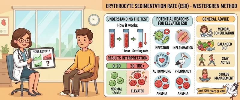
ESR is one of the oldest laboratory tests — over a hundred years old — and one of the most frequently ordered. Yet it is also among the three most commonly misinterpreted markers: patients worry about any deviation, and clinicians sometimes overestimate its specificity. Let's look at what ESR actually measures, why the method matters, how to read a result in context, and when an elevated ESR genuinely demands serious attention.
What Is ESR and How Is It Measured
ESR (erythrocyte sedimentation rate) — the speed at which red blood cells settle to the bottom of a tube over 1 hour. Normally, red cells carry a negative charge and repel each other, settling slowly. During inflammation, acute-phase proteins — fibrinogen, immunoglobulins, C-reactive protein — accumulate in the blood. They coat red cells, neutralise their charge, causing them to clump into "rouleaux" (coin-stack formations) and sediment faster.
In other words, ESR is an indirect reflection of inflammatory proteins in the blood, not a direct measurement of inflammation.
Westergren vs Other Methods
The Westergren method — venous blood with citrate in a 200 mm tube — is the WHO international standard and the most widely used method globally. It gives higher values than older methods, especially when ESR is elevated.
Practically important: if you previously tested by a different method and now use Westergren, results cannot be directly compared. Westergren typically reads 5–20 mm/h higher for the same health status.
Normal ESR Values (Westergren)
| Group | Normal range (mm/h) |
|---|---|
| Men under 50 | 0–15 |
| Men over 50 | 0–20 |
| Women under 50 | 0–20 |
| Women over 50 | 0–30 |
| Pregnancy (2nd–3rd trimester) | up to 40–50 |
| Children 1 month – 14 years | 3–15 |
ESR rises predictably with age — due to increasing chronic low-grade inflammation and changes in plasma protein composition. Applying young-adult norms to elderly patients is a diagnostic error.
A simple formula for age-adjusted upper limits:
- Men: age ÷ 2
- Women: (age + 10) ÷ 2
Why ESR Rises: Mechanism and Causes
ESR rises whenever acute-phase proteins accumulate or plasma protein ratios shift. This occurs in a vast number of conditions — which is precisely why ESR is non-specific.
Infectious and Inflammatory Causes
Acute bacterial infections produce moderate ESR elevation — 30–60 mm/h. Viral infections raise ESR far less than bacterial ones. Chronic infections (tuberculosis, osteomyelitis, endocarditis) produce sustained elevation in the range of 40–80 mm/h.
Autoimmune diseases — rheumatoid arthritis, systemic lupus erythematosus, vasculitis — typically cause pronounced and persistent ESR elevation, often above 60 mm/h.
Malignancy
ESR above 100 mm/h is a red flag requiring oncological evaluation. Particularly characteristic of multiple myeloma (paraproteins dramatically accelerate sedimentation), lymphomas, and metastatic disease. Sustained unexplained ESR > 100 mm/h in an adult is an unconditional indication for comprehensive investigation.
Anaemia
In iron deficiency anaemia and other forms of anaemia, fewer red cells are present and they sediment faster — ESR rises. Important: elevated ESR in anaemia does not indicate inflammation; it is a mechanical effect of reduced blood viscosity.
Kidney and Liver Disease
Chronic kidney disease lowers albumin and alters plasma protein ratios — ESR rises. Nephrotic syndrome with massive albumin loss produces particularly high ESR.
Endocrine Disorders
Hypothyroidism is frequently accompanied by moderate ESR elevation — through changes in protein metabolism. ESR falls when thyroid function is restored.
Physiological Causes
Pregnancy (especially second and third trimester), menstruation, the postpartum period, and older age all cause physiological ESR rise without pathology. Obesity also moderately raises ESR through chronic subclinical inflammation.
Why ESR Falls Below Normal
Very low ESR (0–1 mm/h) is less common but also informative. Causes: polycythaemia (excess red cells), sickle cell anaemia, spherocytosis, hypofibrinogenaemia, severe heart failure, cachexia. In sickle cell anaemia, deformed red cells cannot form rouleaux — ESR is abnormally low even in the presence of inflammation.
ESR and CRP: Why They Are Read Together
C-reactive protein (CRP) and ESR both reflect inflammation but differ in their temporal response:
| CRP | ESR | |
|---|---|---|
| Rise begins | 6–12 hours | 24–48 hours |
| Peak | 24–72 hours | 3–5 days |
| Normalisation | 3–7 days | 2–4 weeks |
| Specificity | Higher | Lower |
Practical significance: CRP reacts fast and normalises fast — an excellent acute-phase marker. ESR reflects a longer process and normalises slowly — a useful marker of chronic inflammation and treatment response.
Discordance between ESR and CRP is often more informative than either alone: high ESR with normal CRP — more likely a chronic process or physiological cause; high CRP with normal ESR — acute inflammation in the first hours before ESR has had time to respond.
How to Prepare and What Affects Results
ESR is part of the standard complete blood count. Blood is drawn fasting in the morning. Important: the sample must be processed within 2 hours of collection, otherwise results are unreliable.
Factors that affect ESR: corticosteroids (lower ESR), NSAIDs (slightly lower), oral contraceptives (raise), pregnancy (raise physiologically), time of day (peak in the afternoon).
After an acute infection, ESR normalises slowly — over 2–4 weeks. A "control" test one week after a respiratory infection will always show elevation even in a fully recovered person.
Degrees of Elevation and Their Clinical Significance
| ESR (mm/h) | Degree | Most likely causes |
|---|---|---|
| Up to 20–30 (F), 15–20 (M) | Normal | — |
| 20–40 | Mild elevation | Viral infection, stress, mild inflammation, age |
| 40–70 | Significant elevation | Bacterial infection, autoimmune disease, anaemia |
| 70–100 | Marked elevation | Severe infections, autoimmune disease in flare |
| > 100 | Extreme elevation | Myeloma, lymphoma, severe sepsis — urgent workup |
These are approximate guides, not algorithms. ESR 40 mm/h in a young pregnant woman is normal; the same value in a 40-year-old man with no complaints warrants investigation.
When to Seek Urgent Medical Attention
Immediately: ESR above 100 mm/h for the first time, especially without an obvious cause — serious infection or malignancy must be excluded first.
Within a few days: ESR 40–70 mm/h in a person under 50 without signs of acute infection; rising ESR on serial testing; ESR above normal combined with unexplained weight loss, night sweats, or enlarged lymph nodes.
Summary
ESR is a sensitive but non-specific marker: it signals that something is happening, but not what. Its value lies in trends and in combination with C-reactive protein and the clinical picture. An isolated moderate ESR elevation in an elderly person or pregnant woman is generally not alarming. ESR above 100 mm/h always warrants comprehensive investigation.
This article is for informational purposes only. Interpretation of test results and treatment decisions are the responsibility of a physician.
For informational purposes only
This article is for informational purposes only and does not constitute medical advice, diagnosis, or treatment. Please consult a healthcare professional for medical guidance.