Red Eyes in Dogs: Conjunctivitis, Injury or Glaucoma
Reviewed by the LabReadAI medical team
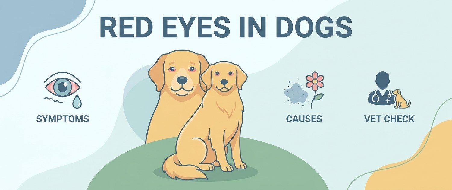
A red eye looks the same across a dozen different diseases, some harmless and some capable of destroying sight within a day. That is exactly why an eye complaint is a poor place for home experiments: the medicine cupboard holds one set of drops while the diagnoses differ, and with some of them the familiar anti-inflammatory drop does irreversible harm. Here is how the main versions look and which signs tell an owner that waiting is not an option.
A Red Eye Is a Symptom, Not a Diagnosis
An eye turns red for two reasons: the superficial conjunctival vessels widen, or the deep vessels around the cornea engorge. The first means surface irritation, the second means inflammation inside the eye or raised pressure. Telling them apart by eye is hard for an owner, but the accompanying signs can be assessed.
Collect a short set of observations:
- one eye or both. A one-sided picture points more towards injury, a foreign body or glaucoma; a two-sided one towards allergy, infection or dry eye;
- pain. The dog squints, rubs with a paw, hides its face, will not let you touch it;
- clarity. A normal cornea is mirror-like and clear; cloudiness, a bluish haze or a white spot is a warning;
- the pupil. Too wide and unresponsive to light, or conversely narrow and irregular;
- discharge. Clear and watery, mucoid, purulent or bloody;
- vision. Does the dog bump into things in unfamiliar surroundings.
Conjunctivitis in Dogs: Common and Overrated
Conjunctivitis is inflammation of the membrane lining the lids and the white of the eye. In dogs it is rarely a disease in its own right: almost always it follows something else — allergy, dryness, irritation, lid anatomy, infection.
How it looks: a red white of the eye, swollen lids, clear or mucoid discharge, moderate discomfort. There is barely any pain, the cornea is clear, the pupil is ordinary and vision is intact.
What usually drives it in a dog:
- atopy and other allergic reactions. In an itchy dog the eyes redden predictably — itching has its own guide, dog scratching;
- irritants — dust, smoke, shampoo, wind through a car window;
- lid anatomy — entropion, misdirected lashes, an excessive fold in flat-faced breeds;
- dry eye — an inadequate tear film;
- infection — usually secondary, on an already damaged surface.
Hence the practical conclusion: treating the conjunctivitis alone without looking for the cause almost always means meeting it again.
Injury and Corneal Ulcer
This is the main reason an eye complaint must not be treated by guesswork. The cornea is the clear front layer, and it is damaged by a branch, another animal's claw, sand, or the dog's own paw during itching.
Signs of a corneal ulcer:
- marked pain: the dog squints, will not open the eye, tears, avoids light;
- heavy tearing, later mucus and pus;
- cloudiness or a bluish haze on the cornea, sometimes a visible pit or spot;
- redness with vessels growing towards the centre in longer-standing cases.
The diagnosis takes a minute with a fluorescein stain: the dye is retained where the epithelium is missing. That is why pain and tearing earn a same-day visit — a deep ulcer progresses to perforation quickly, especially in flat-faced dogs with prominent eyes and incomplete lid closure. What else those breeds pay for their anatomy is in dog breathing heavily.
A cat scratch is doubly dangerous: alongside the mechanical injury it delivers the flora of the animal's mouth and claws, which is covered in Pasteurella multocida.
Entropion and Other Lid Problems
In some breeds the lid margin rolls inward and hair rubs the cornea continuously. That is neither rare nor cosmetic: constant friction produces chronic redness and tearing, and in time ulcers and corneal pigmentation.
What to notice: the eye is red permanently and has been since puppyhood, the tear stain never clears, the dog squints in the wind, and "conjunctivitis treatment" helps only briefly. That picture belongs with an ophthalmologist, because the answer is anatomical rather than pharmaceutical.
The same group holds misdirected lashes, growths on the lid margin, and the prolapsed third eyelid gland seen in young dogs of several breeds.
Keratoconjunctivitis Sicca: Dry Eye
The tear film does more than wet the surface — it feeds and protects the cornea. When there is too little of it, the eye becomes chronically red and the discharge thick and stringy.
Recognisable features of dry eye:
- thick, ropey mucus stuck to the lids while the eye itself looks dull and dry;
- the cornea loses its shine and over time clouds and pigments;
- the picture is bilateral and chronic, with brief "improvement" on any drops;
- commoner in predisposed breeds and in dogs with endocrine disease.
It is checked with a simple Schirmer strip placed under the lid for a minute. That test costs almost nothing and answers a question that otherwise gets answered wrongly for years.
Glaucoma: Here the Clock Runs in Hours
⚠ This is the most urgent of eye situations. In glaucoma the outflow of fluid inside the eye is blocked, the pressure rises and the optic nerve is damaged irreversibly. In dogs irreversible blindness can develop within hours to a day of an acute attack.
What acute glaucoma looks like:
- severe pain: the dog is flat, refuses food, hides, cries when its head is touched — how to read pain behaviour is in dog whining;
- the eye looks engorged, the white is bright red with large tortuous vessels;
- the cornea clouds to a matt blue tone;
- the pupil is wide and does not respond to light;
- the globe feels firmer than the other one and enlarges over time;
- sight in that eye is lost.
The key combination for an owner is severe pain plus cloudiness plus a wide pupil. When that is present, drive in immediately, at night included: measuring the pressure takes seconds, and lost time does not come back.
Uveitis: Inflammation Inside the Eye
Uveitis is inflammation of the vascular layer, and it often turns out to be not an eye disease at all but a window into the body. The pupil behaves in the opposite way to glaucoma: it is narrow, the eye is painful, the cornea may cloud, and haze or blood can be visible inside.
The causes lead far beyond the eye: infections including tick-borne ones, immune disease, tumours, injury, high blood pressure. So with uveitis a vet takes blood and examines the whole animal — how to read the counts and biochemistry is in dog blood test. One specific pairing is diabetes with a rapidly clouding lens: such a dog usually also drinks heavily, and how to measure that is in dog drinking a lot.
Telling an Ordinary Conjunctivitis From an Emergency
| Sign | More like conjunctivitis | More like an emergency |
|---|---|---|
| Pain | Mild, the eye stays open | Severe, the eye is screwed shut |
| Cornea | Clear and shiny | Cloudy, bluish, with a spot |
| Pupil | Ordinary, reacts to light | Wide and fixed, or narrow and irregular |
| Speed | Slow, over days | Within hours, sharply worse |
| Vision | Intact | The dog bumps into things |
| Other eye | Often also red | Usually one eye only |
The rule is simple: pain plus cloudiness plus an altered pupil is not conjunctivitis, whatever it may look like.
When Red Eyes Need an Urgent Vet Visit
- the eye is painful and the dog keeps it shut or half shut;
- the cornea has clouded, or a white, grey or blue spot has appeared;
- the pupil is dilated and does not react to light, the globe feels firm;
- vision has deteriorated sharply and the animal bumps into furniture;
- there was a mechanical event: a branch, claws, a fight, a chemical splash;
- the globe protrudes from the socket after trauma;
- the redness worsens within hours, or there is blood inside the eye;
- red eyes together with lethargy, a temperature and refusing food — how that is worked through is in lethargic dog.
What Not to Do at Home: Drops From the Family Cabinet
- Do not use human eye drops, steroid ones above all: with a corneal ulcer they suppress healing and promote tissue breakdown, and the eye can be lost. Anything that goes into an eye is prescribed by a vet after an examination.
- Do not use vasoconstrictor drops "to take the redness away": they mask the very sign the diagnosis rests on.
- Do not rinse the eye with tea, herbal infusions or anything containing alcohol.
- Do not try to remove a foreign body from under the lid yourself with tweezers or a cotton bud.
- Do not give human painkillers: they are dangerous for dogs, and eye pain is an important diagnostic sign.
- Do not postpone the visit when the pain is severe: an eye has a very short margin.
- Do not remove a protective collar prescribed after an injury: with one paw a dog turns a surface wound into a full-thickness one in an evening.
Which Examinations Will Clarify the Redness
An ophthalmic consultation consists almost entirely of quick tests:
- fluorescein staining — is there a corneal defect;
- intraocular pressure measurement — glaucoma or uveitis;
- a Schirmer test — is there enough tear fluid;
- slit lamp examination and ophthalmoscopy — the front of the eye, the lens, the fundus;
- cytology and culture — for purulent discharge and non-healing ulcers;
- blood count, biochemistry, blood pressure, infectious disease tests — for uveitis and bilateral change;
- ocular ultrasound — when the media are opaque and you cannot look inside: what ultrasound adds is in ultrasound for dogs and cats;
- CT or MRI of the orbit — for a protruding eye, a suspected abscess or a mass behind the globe; how the methods differ is in MRI and CT for pets.
In cats a red, watering eye reads differently: upper respiratory viral infection comes first there, as covered in cat watery eye and cat sneezing. In rabbits and rodents tearing and redness usually trace back to the teeth rather than the eye: see small pet diseases.
A finished form can go into pet results interpretation and an imaging report into pet imaging interpretation: the service explains the wording in plain language and builds questions for the vet. If there are no studies yet, describe your symptoms and browse dog symptoms.
This article is informational. Diagnosis and treatment of your pet are the job of a veterinarian.
For informational purposes only
This article is for informational purposes only and does not constitute medical advice, diagnosis, or treatment. Please consult a healthcare professional for medical guidance.