Melatonin Blood Test: Normal Levels, Function and Causes
Reviewed by the LabReadAI medical team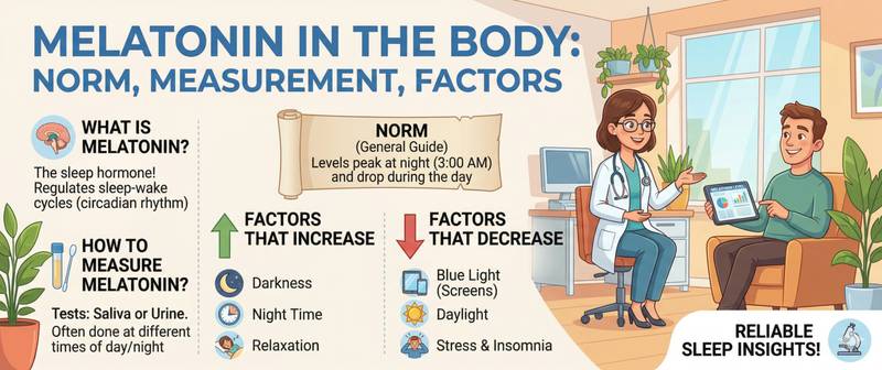
Melatonin is called the "hormone of darkness" — and that is an accurate description: it is produced only at night, strictly in the absence of light, and its daily oscillations signal to every cell in the body that it is nighttime and time to recover. Most people encounter melatonin through sleep supplement advertising, but as a diagnostic marker it is considerably more interesting and complex. Let's break down what it actually reflects, how to measure it correctly, and what deviations mean.
What Melatonin Is and How It Is Produced
Melatonin is an indolamine hormone synthesized by the pineal gland (epiphysis) from tryptophan via serotonin. Synthesis is strictly light-dependent: photoreceptors in the retina detect light and suppress pineal activity via the retinohypothalamic tract. In darkness this inhibition is removed — melatonin production begins approximately 2 hours after dark onset, peaks at 2–4 AM, and falls to near-zero by 6–8 AM.
Melatonin serves several core functions. Above all, it is the principal "zeitgeber" — the circadian rhythm setter — synchronizing the internal biological clock with the external light-dark cycle. Its nocturnal peak signals every organ and tissue to enter "night mode": body temperature drops, metabolism slows, and cellular repair processes intensify.
Beyond sleep regulation, melatonin is a potent antioxidant, participates in immune regulation, suppresses gonadotropin secretion in children (protecting the reproductive axis from premature activation), and exerts oncostatic effects that are being actively investigated.
A full discussion of melatonin physiology and clinical applications is in the article melatonin: what it is and how to fix your sleep.
Melatonin is not synthesized exclusively in the pineal gland — intestinal cells, the retina, skin, and immune cells also produce it. But only pineal melatonin circulates in diagnostically relevant quantities and delivers the systemic chronobiological signal.
Normal Melatonin Levels: Blood and Urine
Melatonin has fundamentally different levels at different times of day. This is why "a normal melatonin value" stated without time of collection is biologically meaningless.
In blood (serum/plasma):
| Time of day | Normal in adults (pg/mL) |
|---|---|
| Daytime (8:00 AM–8:00 PM) | 1–10 |
| Early evening (8:00–11:00 PM) | 20–60 |
| Night (11:00 PM–3:00 AM) | 80–200 |
| Peak (2:00–4:00 AM) | 100–300 |
| Morning (6:00–8:00 AM) | < 20 |
In urine — 6-sulfatoxymelatonin (6-SOMT), the principal melatonin metabolite reflecting total nocturnal melatonin output:
| Group | Normal 6-SOMT (nmol/24h) |
|---|---|
| Adults 18–40 years | 20–50 |
| Adults 40–65 years | 15–40 |
| Elderly over 65 years | 5–25 |
In pre-pubertal children, melatonin levels are substantially higher than in adults — one of the reasons children sleep deeply and for long periods. At puberty, melatonin falls under the influence of sex hormones. Levels continue to decline with age: by 60–70 years, the nocturnal peak in many people is half what it was in young adulthood.
During pregnancy, melatonin physiologically rises — particularly in the third trimester, where it participates in programming the fetal circadian rhythm.
How to Collect Samples for Melatonin Testing
A melatonin test is one of the most technically demanding in endocrinology. Errors in sample collection completely invalidate the result.
Three main testing approaches:
1. Blood melatonin — single time-point draws or serial nocturnal sampling. Conditions are stricter than for any other hormone:
- Blood must be drawn in complete darkness or under red light (wavelength > 600 nm): any white light immediately suppresses secretion
- Nocturnal draw: 2:00–4:00 AM, provided the patient went to bed no later than 11:00 PM and did not wake in the light
- Daytime draw (for comparison): 10:00 AM–12:00 PM under standard conditions
- No alcohol or caffeine for 48 hours
- Sample must be frozen immediately — melatonin is unstable at room temperature
2. Overnight urine for 6-SOMT — technically simpler and more practical for routine clinical use:
- Collect overnight urine (10:00 PM to 8:00 AM the next morning) or full 24-hour urine
- Keep refrigerated throughout the collection period; add preservative (acetic acid)
- No lighting restrictions — more convenient for outpatient patients
3. Saliva — the preferred method for research and for measuring the dim-light melatonin onset time (DLMO). Less commonly used in routine clinical practice.
Important: blood melatonin levels do not reflect biological activity in tissues — receptor sensitivity to melatonin is individually variable. A normal blood level in a patient with insomnia does not exclude chronobiological pathology.
Causes of Low Melatonin
Low melatonin is a far more common clinical problem than elevated melatonin. Modern lifestyle is one of the most powerful suppressors of melatonin secretion.
Light pollution and screens. Blue light at 460–480 nm (smartphone and laptop screens, LED lighting) is maximally effective at suppressing melatonin through melanopsin receptors in the retina. Screen use at night delays the melatonin onset by 1.5–3 hours — effectively phase-shifting the entire circadian rhythm.
Age. Physiological decline in pineal function is a key mechanism of insomnia in older adults. Progressive pineal calcification reduces the number of active pinealocytes. By age 70, the nocturnal melatonin peak is on average 2–4 times lower than at 25–30 years.
Night shifts and irregular schedules. Night work completely inverts or destroys the normal melatonin profile. Chronic circadian disruption in shift workers is associated with elevated risks of breast cancer, metabolic syndrome, and depression.
Cortisol and chronic stress. Cortisol and melatonin are physiological antagonists with reciprocal daily rhythms: when one is high, the other is suppressed. Chronically elevated cortisol suppresses melatonin synthesis via GABAergic and noradrenergic mechanisms.
Melatonin-lowering medications: beta-blockers (suppress noradrenergic pineal stimulation — one of the most thoroughly studied drug effects on melatonin), NSAIDs, chronic benzodiazepine use, and some antidepressants.
Tryptophan and vitamin B6 deficiency — reduced substrate for serotonin → melatonin synthesis.
Pineal tumors and trauma — rare but direct causes of hypomelatoninemia.
Causes of Elevated Melatonin
Significant melatonin elevation is less common and has a narrower clinical significance in routine practice.
Pineal tumors (pineocytomas/pineoblastomas). Secreting pineal tumors in children can produce marked hypermela-toninemia. Clinically, this manifests as delayed puberty — high melatonin suppresses the gonadotropic axis. Non-secreting pineal tumors, conversely, lower melatonin by destroying gland tissue.
Blindness. In patients with total light imperception, the circadian melatonin rhythm cannot synchronize with the external light-dark cycle — the rhythm becomes "free-running," often resulting in melatonin elevations during daytime hours.
Psychiatric conditions. Abnormally elevated melatonin is found in some forms of depression and in bipolar disorder; seasonal affective disorder (SAD) is associated with the opposite — reduced melatonin amplitude. Chronobiological dysregulation is part of the pathophysiology of depression.
Thyroid dysfunction. In hypothyroidism, reduced hepatic melatonin catabolism can lead to level accumulation.
Exogenous melatonin supplementation — obviously elevates measured levels and invalidates diagnostic testing. Testing while taking melatonin supplements is uninformative.
Melatonin and Other Hormones: Systemic Interactions
Melatonin does not exist in isolation — it is embedded in a complex hormonal network, and its deviation frequently coexists with changes in other markers.
Melatonin and cortisol — the reciprocal axis. The normal daily rhythm: melatonin peaks at 2–4 AM → falls to near-zero by 6–8 AM → at this precise moment cortisol surges, driving awakening. Under chronic stress, cortisol fails to fall by evening — and melatonin cannot rise normally. Normalizing cortisol through stress management and sleep regularity restores the melatonin rhythm far more effectively than supplementation alone. The mechanism is detailed in how to lower cortisol in women.
Melatonin and TSH. Melatonin exerts an inhibitory influence on TSH secretion and thyroid hormone output — via receptors in the hypothalamus and pituitary. Chronic nocturnal melatonin deficiency may allow the thyroid axis to run in "accelerated mode." This is one proposed mechanism explaining why sleep disorders and subclinical hyperthyroidism frequently coexist.
Melatonin and prolactin. Both hormones have a nocturnal peak and both are suppressed by light. Melatonin stimulates prolactin secretion through hypothalamic receptors — under a normal circadian rhythm their nocturnal peaks are synchronized. Disruption of this synchrony in night-shift workers is one mechanism explaining menstrual irregularities in this population.
Melatonin and the reproductive system. In children, high melatonin suppresses premature pubertal activation. In adults, seasonal melatonin variations (longer nights = more melatonin) modulate reproductive activity in seasonally breeding mammals — in humans this effect is minor but detectable in seasonal testosterone patterns.
When Melatonin Should Be Tested and Who to See
Melatonin measurement is not a routine screening test. It is clinically meaningful in specific situations and is typically ordered by a specialist — sleep medicine physician, neurologist, endocrinologist, or chronobiologist.
Indications for melatonin testing:
- Chronic insomnia not responding to standard treatment — to assess the chronobiological component
- Circadian rhythm disorders: delayed sleep phase syndrome (DSPS), advanced sleep phase syndrome, irregular sleep-wake disorder
- Night shift work with pronounced sleep and metabolic disturbances
- Delayed or precocious puberty in children
- Suspected pineal tumor (in conjunction with brain MRI)
- Monitoring chronobiological treatment effectiveness (light therapy, melatonin)
- Seasonal affective disorder with depressive episodes during dark months
Self-prescribing melatonin supplements without measuring levels and assessing circadian timing is frequently ineffective or counterproductive when taken at the wrong time. The question "when to take melatonin" for an individual patient requires knowing their personal DLMO (dim-light melatonin onset) — which can only be established through laboratory testing.
This article is for informational purposes only and does not replace professional medical advice. Consult a sleep specialist, neurologist, or endocrinologist for sleep disturbances.
For informational purposes only
This article is for informational purposes only and does not constitute medical advice, diagnosis, or treatment. Please consult a healthcare professional for medical guidance.