Elevated ESR: Causes, Normal Range and Blood Test Results
Reviewed by the LabReadAI medical team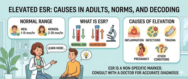
Your blood test comes back and the ESR line is flagged high. Or your doctor mentions "raised ESR" as a reason for further investigation — even though you feel perfectly well. Erythrocyte sedimentation rate is one of the oldest and simultaneously one of the least specific markers in laboratory medicine. That is precisely the point: a high ESR says that "something may be wrong" but does not say what. This article covers reference ranges, causes, and how to correctly interpret this result.
What Is ESR and Why Is It Tested
ESR stands for erythrocyte sedimentation rate. When blood is placed with an anticoagulant in a vertical tube and left to stand for an hour, red blood cells settle to the bottom under gravity — forming a red column below and a clear plasma layer above. The rate at which they settle, measured in millimetres per hour, is the ESR.
Why does ESR change with illness? Red blood cells carry a negative surface charge and normally repel each other, settling slowly. During inflammation, acute-phase proteins accumulate in blood plasma: fibrinogen, immunoglobulins, α-2-globulins. These proteins coat red blood cells, neutralise their negative charge, and promote the formation of "rouleaux" — stacked chains of cells that settle significantly faster. This is why ESR is an indirect marker of inflammation, not a direct measure of it.
ESR is part of the complete blood count and is one of the most frequently ordered tests in outpatient medicine. It does not point to a specific disease but acts as a sensitive alarm: a significantly elevated ESR without an obvious cause is grounds for targeted investigation.
An important limitation: ESR is a slow marker. It begins to rise 24–48 hours after inflammation starts and normalises with a similar delay — sometimes weeks after recovery. For assessing acute, immediate inflammation, C-reactive protein is far more accurate: it responds within 6–12 hours and falls equally quickly when treatment is effective.
How to Prepare for an ESR Test
ESR is measured in venous or capillary blood as part of a complete blood count. Preparation is minimal, but ignoring the basic rules can distort the result.
Timing. Preferably in the morning, between 7 and 11 am. ESR shows small circadian variation — it is marginally higher in the afternoon. For serial monitoring, always test at the same time of day.
Food. A fatty meal the evening before raises plasma chylomicron levels, which — like inflammatory proteins — accelerate red cell sedimentation. A light dinner and a 4–8-hour fast before the draw is recommended; water is unrestricted.
Exercise and stress. Intense exercise the day before and acute stress transiently elevate ESR. Avoid both for 24 hours before the test.
Infections and vaccination. Even a mild respiratory infection raises ESR for several weeks. For a planned test, postpone until 3–4 weeks after full recovery. Vaccination also produces a brief elevation.
Methods. The Westergren method is the international standard. The Panchenkov method is used in some countries and gives comparable but not fully interchangeable results. When comparing serial measurements, ensure the same method is used at the same laboratory throughout.
ESR Normal Range by Age and Sex
ESR varies substantially with sex and age. Reference values are higher in women than men — partly due to lower haematocrit and the influence of sex hormones on plasma protein composition. The physiological normal range rises gradually with age.
| Group | ESR Normal Range (Westergren) | Notes |
|---|---|---|
| Men under 50 | 0–15 mm/h | — |
| Men over 50 | 0–20 mm/h | Physiological rise |
| Women under 50 | 0–20 mm/h | — |
| Women over 50 | 0–30 mm/h | Physiological rise |
| Pregnant women | Up to 45–50 mm/h | Physiologically normal |
| Children under 12 | 2–12 mm/h | Age-dependent |
Clinical significance thresholds:
- 20–40 mm/h — mild elevation: context-dependent; often physiological or minor inflammation.
- 40–70 mm/h — marked elevation: active inflammation, autoimmune process, or infection.
- > 100 mm/h — significantly accelerated: serious organic pathology; source must be found promptly.
The key interpretive rule: an isolated elevated ESR with a normal full blood count, normal CRP, and no symptoms warrants a repeat test in 2–4 weeks — not an immediate oncology workup.
Causes of Elevated ESR: Inflammation, Infection and Beyond
ESR rises in any condition that alters plasma protein composition or red cell morphology. The list of causes is very broad — which is precisely why the test lacks specificity.
Autoimmune and Inflammatory Diseases
The most common cause of high ESR in rheumatological practice. Rheumatoid arthritis — ESR in active disease typically reaches 40–80 mm/h and correlates with the DAS28 disease activity index. Falling ESR on treatment is a reliable marker of therapeutic response. Systemic lupus erythematosus, dermatomyositis, and systemic sclerosis — ESR rises predictably during flares. Inflammatory bowel disease (Crohn's disease, ulcerative colitis) produces moderate-to-marked ESR acceleration in the active phase.
For all autoimmune processes, ESR is assessed alongside rheumatoid factor and other disease-specific markers — never in isolation.
Infections
Acute bacterial infections — pneumonia, pyelonephritis, abscesses, bacterial endocarditis — produce a rapid, substantial rise in ESR, frequently above 60–80 mm/h. Viral infections elevate ESR more modestly. Infectious mononucleosis is a notable exception — it can markedly elevate ESR alongside a rise in atypical lymphocytes in the blood count.
Chronic infections — tuberculosis, chronic hepatitis, autoimmune hepatitis — cause persistently moderate-to-marked ESR acceleration, often discovered incidentally on routine testing.
Anaemia and Other Causes
Iron deficiency anaemia is one of the most common causes of moderately elevated ESR in the absence of inflammation. The mechanism: fewer red cells per unit volume reduces the physical opposition to sedimentation. The distinguishing feature: CRP is normal in pure iron deficiency anaemia.
Pregnancy is a physiological cause of high ESR requiring no further investigation: values up to 45–50 mm/h in the third trimester are normal and resolve within 3–6 weeks of delivery.
Malignancies — particularly lymphomas, multiple myeloma, renal cell carcinoma, and ovarian cancer — frequently present with persistently elevated ESR without obvious symptoms. In multiple myeloma, ESR can exceed 100 mm/h due to massive paraprotein production.
Other causes: renal failure (accumulation of uraemic proteins), hypothyroidism, obesity, and smoking.
Very High ESR: What Does a Result Above 100 mm/h Mean
ESR above 100 mm/h is not simply "elevated" — it is a clinical situation that almost always indicates serious organic pathology and requires immediate investigation.
Statistically, the causes of ESR > 100 mm/h break down as follows: approximately 35% — infections (most commonly severe bacterial: tuberculosis, bacterial endocarditis, deep abscesses); approximately 25% — malignancies (particularly haematological — lymphomas, multiple myeloma); approximately 20% — systemic autoimmune diseases (vasculitis, polymyalgia rheumatica, Sjögren's syndrome); the remainder — renal failure and severe chronic disease.
When ESR exceeds 100 mm/h without an established diagnosis, the standard investigation algorithm includes: full blood count with differential; CRP, fibrinogen, serum protein electrophoresis; liver and kidney biochemistry; urinalysis; chest X-ray; tumour marker screen; rheumatological markers. Normal results across all of the above with persistently very high ESR is an indication for haematology referral and PET/CT.
Low ESR: Causes and Clinical Significance
ESR below 2 mm/h in adults is far less common than elevation, and in most cases is not pathological.
Polycythaemia vera — a pathologically elevated haematocrit. The densely packed red cells physically cannot settle quickly — ESR falls to 1–2 mm/h. This is a useful indirect sign of polycythaemia when acute-phase proteins are normal.
Sickle cell anaemia — the abnormal shape of sickle cells prevents rouleaux formation, which is required for sedimentation to occur.
Hyperviscosity states in certain conditions (protein deficiency, cachexia) also reduce ESR.
Technical causes: incorrect blood-to-anticoagulant ratio, delayed processing, or inappropriate temperature during storage. When ESR is unexpectedly very low, the first step is to verify the conditions of sample collection and handling.
An isolated low ESR in the presence of normal other tests in an asymptomatic person is not a cause for concern.
When to See a Doctor Urgently
Seek prompt medical attention in the following situations:
- ESR above 100 mm/h under any circumstances — even without symptoms: this value always requires an active search for the underlying cause.
- Elevated ESR (> 40 mm/h) combined with night sweats, unexplained weight loss, and lymph node enlargement — the classic "B-symptom" triad of lymphoma, warranting haematological evaluation.
- Persistently elevated ESR over 3 or more months without an identified cause — indication for a comprehensive systematic workup.
- ESR rises sharply after a period of normal values without any apparent cause (no infection, no injury) — possible marker of a new inflammatory or neoplastic process.
- High ESR in an older adult (over 60) with pain and stiffness in the shoulder and pelvic girdle — the characteristic picture of polymyalgia rheumatica, which responds rapidly to glucocorticoids when diagnosed promptly.
- Elevated ESR in a patient on immunosuppressive therapy — the immune system may not mount a full response to infection; ESR may be the only laboratory signal of an occult infective process.
This article is for informational purposes only and does not replace medical consultation. Result interpretation and diagnosis are made by a qualified physician.
For informational purposes only
This article is for informational purposes only and does not constitute medical advice, diagnosis, or treatment. Please consult a healthcare professional for medical guidance.