Autoimmune Hepatitis: Symptoms, Diagnosis and Treatment
Reviewed by the LabReadAI medical team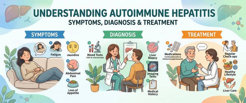
Elevated ALT and AST with no viral hepatitis, no alcohol, and no toxic exposure — this is the classic presentation that frequently leads to a diagnosis of autoimmune hepatitis. In this disease, the immune system mistakenly attacks the body's own liver cells. It progresses slowly but, without treatment, inevitably leads to cirrhosis. The good news: when diagnosed in time, autoimmune hepatitis responds excellently to immunosuppressive therapy and remission is achievable in the vast majority of patients.
What Autoimmune Hepatitis Is and Why It Develops
Autoimmune hepatitis (AIH) is a chronic inflammatory liver disease in which T-lymphocytes and autoantibodies attack hepatocytes, misidentifying them as foreign. The trigger mechanism is not fully understood: a genetic predisposition (HLA-DR3 and HLA-DR4 haplotypes) combined with an environmental trigger — viral infection, medications, or another immune stressor — is believed to initiate the disease.
A defining characteristic of AIH is female predominance: 70–80% of patients are women. The disease can begin at any age but shows two incidence peaks: 10–30 years and after 40 (perimenopausal period).
AIH is classified into two types based on the serological profile. Type 1 — the most common (80% of cases): antinuclear antibodies (ANA) and/or anti-smooth muscle antibodies (ASMA). Type 2 — less common, more frequent in children: anti-liver-kidney microsome type 1 antibodies (anti-LKM-1). Typing has no practical bearing on treatment selection but helps confirm the diagnosis.
Symptoms of Autoimmune Hepatitis
AIH is a master of disguise. Approximately one third of patients are diagnosed incidentally — during a routine biochemical check with no symptoms whatsoever.
Symptoms of chronic disease (the most common presentation):
- Fatigue and reduced work capacity — the most frequent complaint
- Discomfort or heaviness in the right upper quadrant
- Mild scleral icterus
- Arthralgia — joint pain without overt inflammation
- Acne and menstrual irregularities in women (related to hormonal changes secondary to liver disease)
Acute onset — in 25–30% of patients: the picture resembles acute hepatitis with pronounced jaundice, dark urine, and pale stools. Occasionally it debuts as fulminant hepatic failure — rare but severe.
Extra-hepatic manifestations — a hallmark of AIH: thyroiditis, rheumatoid arthritis, coeliac disease, inflammatory bowel disease. Their combination with elevated transaminases is a strong argument for an autoimmune aetiology.
Diagnosis: Which Tests Confirm the Diagnosis
AIH diagnosis is composite — established from a combination of biochemical, serological, and histological findings. The simplified IAIHG scoring system is used in practice.
ALT and AST — hepatocyte injury markers. In AIH they are typically moderately or significantly elevated (2–10 times the upper limit of normal); extreme elevations, as in acute viral hepatitis, are less common. An AST/ALT ratio < 1 is typical of active inflammation; > 2 suggests alcoholic liver disease.
Gamma-globulins and IgG — AIH characteristically causes a substantial rise in serum immunoglobulin G (IgG): > 2× the upper limit of normal. This is one of the key diagnostic criteria. Total protein may be normal or elevated.
Bilirubin — elevated with active inflammation and decompensation. Normal bilirubin does not exclude active AIH.
Albumin and prothrombin time — markers of hepatic synthetic function. Falling albumin and prolonged PT indicate advanced disease or acute decompensation.
GGT and ALP — mildly elevated in AIH. A markedly elevated ALP alongside only moderately raised transaminases points more toward primary biliary cholangitis or primary sclerosing cholangitis — important differential diagnoses.
CRP — typically mildly elevated or normal in AIH. A very high CRP suggests a bacterial complication rather than autoimmune disease activity.
Autoantibodies — the serological cornerstone:
- ANA (antinuclear antibodies) — positive in type 1 AIH
- ASMA (anti-smooth muscle antibodies) — more specific than ANA for type 1
- Anti-LKM-1 — the type 2 AIH marker
- Anti-SLA/LP (anti-soluble liver antigen) — highly specific for AIH
Liver biopsy — required for definitive diagnosis and fibrosis staging. The characteristic histological pattern: interface hepatitis (periportal hepatitis) with plasma cell infiltrates. Elastography (FibroScan) is a less invasive alternative for fibrosis monitoring.
Comprehensive liver assessment uses the liver function panel — the mandatory starting point for any suspected liver disease.
Differential Diagnosis: What Must Be Excluded
Before diagnosing AIH, other causes of chronic hepatitis must be ruled out: viral hepatitis B and C, alcoholic hepatitis, non-alcoholic steatohepatitis, drug-induced liver injury (DILI), Wilson's disease, and haemochromatosis. Distinguishing AIH from DILI is particularly important — they are clinically identical, but management differs fundamentally.
Primary biliary cholangitis (PBC) and primary sclerosing cholangitis (PSC) can coexist with AIH — this is called "variant syndrome" or "overlap syndrome" and requires a specific therapeutic approach.
Treatment of Autoimmune Hepatitis
AIH responds well to immunosuppressive therapy — it is one of the few chronic liver diseases where medical treatment fundamentally changes the prognosis.
First line — prednisolone + azathioprine:
- Prednisolone 40–60 mg/day initially, tapered to a maintenance dose of 5–10 mg/day
- Azathioprine 50–150 mg/day is added after 2 weeks to allow steroid dose reduction and maintain remission
- Goal: normalisation of ALT, AST and IgG + histological remission
Alternative agents for intolerance or inadequate response: budesonide (fewer systemic side effects, but only when cirrhosis is absent), mycophenolate mofetil, tacrolimus.
Duration of treatment — minimum 2–3 years, in most cases lifelong: after withdrawal, relapse occurs in 80% of patients within 3 years.
Liver transplantation — for decompensated cirrhosis or fulminant hepatic failure not responding to therapy. AIH can recur in the transplanted organ.
Connection with Other Autoimmune Diseases
AIH rarely exists in isolation. In 30–40% of patients, other autoimmune diseases are found simultaneously: Hashimoto's thyroiditis is the most common co-diagnosis, followed by rheumatoid arthritis and inflammatory bowel disease. This means that at the time of AIH diagnosis, thyroid function should be checked and screening for other autoimmune conditions considered.
When to Seek Urgent Medical Attention
Call emergency services or go to hospital immediately for: worsening jaundice with dark urine and pale stools; ascites — abdominal distension and dyspnoea; confusion or encephalopathy; sudden deterioration in a known AIH patient in the context of infection.
See a hepatologist or gastroenterologist within 1–2 weeks for: ALT and AST elevated 3–5 times the upper limit of normal without an obvious cause; elevated IgG combined with abnormal transaminases; positive ANA or ASMA on routine testing.
This article is for informational purposes only. Diagnosis and treatment of autoimmune hepatitis are carried out by a hepatologist or gastroenterologist.
For informational purposes only
This article is for informational purposes only and does not constitute medical advice, diagnosis, or treatment. Please consult a healthcare professional for medical guidance.