Enlarged Lymph Nodes in Children: When It Is Normal
Reviewed by the LabReadAI medical team
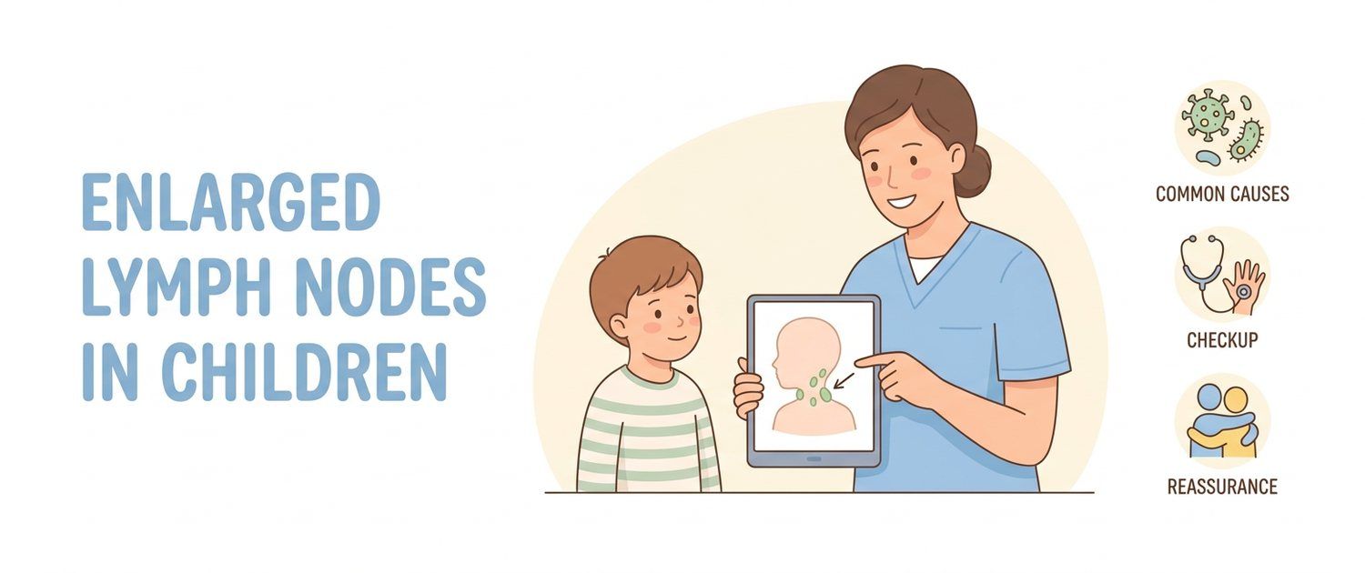
A parent feels a lump on a child's neck and immediately thinks of the worst. The statistics point the other way: in children enlarged lymph nodes are almost always reactive, reflecting the normal work of the immune system. The task is not to panic and yet not to miss the few features that genuinely need attention.
Enlarged lymph nodes in children: why it is usually normal
A lymph node is a filter for lymph draining its own territory. In children lymphoid tissue is physiologically active: it meets most infections for the first time and responds by enlarging. So palpable nodes in a child are the rule rather than the exception, particularly between ages 2 and 10.
Reactive enlargement develops over days and resolves over weeks. A parent who palpates the node daily and sees no change in a week often concludes it "is not going away" — although that is the ordinary pace.
Lymph nodes in a child's neck: sizes and what counts as enlarged
| Location | Normal in children | Considered enlarged |
|---|---|---|
| Cervical, axillary | Up to 1 cm | Over 1 cm, especially if growing |
| Inguinal | Up to 1.5 cm | Over 1.5 cm |
| Occipital, post-auricular | Up to 1 cm, common in infants | Over 1 cm after age 2 |
| Supraclavicular | Not palpable normally | Any palpable node |
Besides size, four properties are assessed: mobility (rolling freely under the fingers is good), consistency (soft-elastic good, stony hard not), tenderness (a tender node is more often inflammatory; a painless hard one is more concerning) and fixation to skin or to each other.
Lymph nodes behind the ear and at the back of the head
Post-auricular and occipital nodes respond to anything happening on the scalp: seborrhoeic dermatitis, bites, small wounds, head lice, ear infection. In infants occipital nodes are often palpable and mean nothing. A classic exception is occipital enlargement in rubella, see measles, rubella and mumps.
Lymphadenitis in children: inflammation of the node itself
Lymphadenitis means the node itself is inflamed rather than reacting to a distant infection. The picture differs sharply: the node grows rapidly within a day or two, becomes painful, the overlying skin turns red and hot, and fever appears. Usually this is a bacterial infection spreading from tonsils, teeth or a skin wound; a common entry point is tonsillitis, see tonsillitis.
This state needs a doctor: it is treatable, and if neglected the node can suppurate.
Swollen nodes without fever: what is considered
Absent fever makes an acute bacterial cause unlikely. Then the possibilities are:
- residual reactive enlargement after a past infection — the commonest;
- cat scratch disease — a node on the arm or in the armpit 1–3 weeks after a scratch or bite;
- toxoplasmosis — usually cervical nodes, see toxoplasmosis;
- atypical mycobacteria — a firm painless node in a preschooler with violaceous overlying skin;
- tuberculosis — in a protracted course, see tuberculosis and the skin tests described in Mantoux and Diaskintest.
Lymph nodes in mononucleosis and other infections
In infectious mononucleosis the picture is recognisable: symmetrically enlarged cervical and posterior cervical nodes, tonsillitis, a blocked nose without discharge, marked fatigue and often an enlarged liver and spleen. Atypical mononuclear cells appear in the blood. The culprit is Epstein–Barr virus, whose family is described in herpes virus types.
Prolonged fever with node enlargement and no obvious focus is covered separately in fever without symptoms.
When lymph nodes in children are dangerous
Vigilance is warranted with any of these:
- a supraclavicular node of any size;
- a node over 2 cm that keeps growing beyond two weeks;
- a node that is hard, painless and fixed to surrounding tissue;
- generalised enlargement: nodes in three or more non-adjacent regions;
- accompanying weight loss, night sweats, fever lasting over a week;
- pallor, bruising, bleeding — a possible blood-forming problem;
- an enlarged liver and spleen;
- a node that has not shrunk within 4–6 weeks of observation.
Which tests are done for enlarged lymph nodes
A typical reactive node after a viral illness needs observation, not tests. Investigation follows warning signs or a protracted course:
- a complete blood count with differential is the key first step; paediatric ranges are in CBC in children, and the behaviour of lymphocytes in viral infection explains much of the picture;
- CRP and ESR as inflammation markers, see also raised ESR;
- Epstein–Barr virus antibodies — the EBV marker;
- tuberculin skin tests when a tuberculous cause is suspected;
- ultrasound of the node, assessing structure, blood flow and margins, often settling the question without biopsy;
- biopsy only on a doctor's decision when concern persists.
This article is informational. Lymph nodes are assessed by a doctor on examination.
For informational purposes only
This article is for informational purposes only and does not constitute medical advice, diagnosis, or treatment. Please consult a healthcare professional for medical guidance.